Jul 112016
 

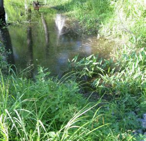
There’s a natural pond down the road, where I collect samples. I always go to the same spot, the edge of a shallow basin in the “littoral zone”, where the pond shades into the shore. I follow the same routine each time: I dip a turkey baster into the soft muck–organic sediments enriched with duckshit and decomposing leaves–and suck out a few cc’s for the bottom of the jar; then I ladle a bit of surface water over the mud, to create a miniature approximation of the natural site.

Turtle Pond2

Sampling Site in Turtle Pond

I’ve taken hundreds of samples there over the years, from an area no bigger than a coffee table.  And, despite having done this regularly for about half a decade, nearly every jar I bring home contains something I’ve never seen before.

Of course, to a microbe, a coffee table-sized site is actually quite big. If you were a typical one-celled protist–say, 50 micrometres from tip to tail–a four-foot patch of pond would be more than 24,000 times longer than your body. From a microbe’s point of view, then, my sampling basin is equivalent to a lake 28 miles long. Scaled up to human size, the entire pond would be at least 1,350 miles across, with a greater surface area than the Mediterranean Sea! So, there’s quite a lot of available habitat in the small body of water I call “Turtle Pond.”

At any given time, only a small fraction of the site’s potential diversity will be visible. The mix of species can change from one hour to the next, as weather and random events alter water chemistry, light levels, temperature and organic nutrients. A few hours of sunlight might kick off a sudden bloom of cyanobacteria, soon followed by nassulid ciliates that eat strands of blue algae like spaghetti; then, a few minutes of rain disperse the bloom, the nassulids disappear, and are replaced by some other combination of species. The new populations have barely established themselves, when a turtle (which, from a microbe’s point of view, is about 7 miles long), blunders into the shallows, churning up mud, mixing oxygen-rich upper layers with the anoxic sediments below, and everything changes again. Each shift in the pond creates new opportunities for some organisms, and makes life impossible for others. One species blooms, and another goes into dormancy, withdrawing into resting cysts or simply lurking in reduced numbers, waiting for better days.

The microbial “seed bank” can store a staggering diversity of such hidden opportunists, which emerge from their cysts only when conditions are exactly right. Some are “weed” species, generalists that crop up in nearly every sample. Others have very particular needs, and rarely appear. A few may never find what they need to grow and reproduce. If any cysts of salt-loving (halophilic) protists should blow in from some distant shore, they are probably out of luck.

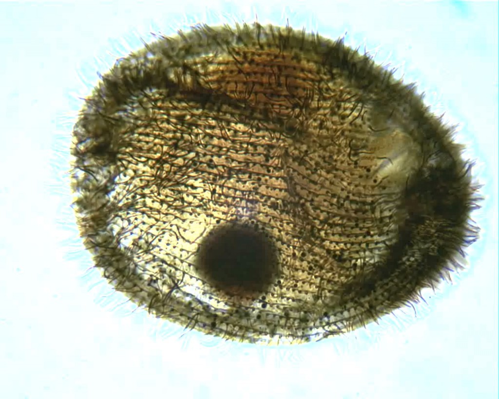
Recently, I brought home a few jars from the usual spot, and found three rather flamboyant species to cross of my life list. One was Actinobolina radians, an oddball I’ve been hoping to find for years (ever since I first saw it’s mugshot in Vladimir Schewiakoff’s beautifully-illustrated Infusoria Aspirotricha of 1889). Actinobolina is a ciliate in the class Litostomatea, a group of rapacious and morphologically hilarious carnivores. The class includes such celebrity ciliates as the improbably elastic Lacrymaria, the elephant-nosed Dileptus, and a frisky little keg full of death called Didinium, which eats other ciliates like popcorn. Like its cousins, Actinobolina is a hunter. However, it has a unique strategy for capturing prey. When swimming about, it looks much like any other ciliate: egg-shaped, and uniformly covered with cilia. Once it has a found a suitable feeding site, it comes to a stop and slowly extends a large number of long, stiff tentacles, each of which carries toxic extrusomes (cellular darts) to be released on contact into its victim’s body. In this state, it looks rather like a heliozoan “sun animalcule”.   With its tentacles extended, Actinobolina lies in wait like a spider in its web, until some tasty creature–a rotifer , or another ciliate–bumps into it. The toxic extrusomes are deployed and the victim is paralyzed, whereupon the ciliate withdraws its tentacles, pulling the stunned prey toward its cellular mouth.

Unfortunately, I didn’t manage to record a successful hunt, but I did get some footage of the ciliate extending and retracting its tentacles.  And really, I was just pleased to have seen this bizarre organism.

Another creature that appeared in my jars was the odd, oxygen-hating amoebozoan Pelomyxa, an indiscriminately hungry thing that looks like a tumbling bag full of rocks and food. I see Pelomyxa fairly often, but this one was a different species, smaller and more colorful, and with a distinctive style of movement, featuring frequent lateral “eruptions” of pseudopods. I believe it is Pelomyxa binucleata, a species first identified in the 19th century. For a time, in the late 20th century, it was believed that P. binucleata and all “species” of Pelomyxa, were really just forms and phases of a single species, Pelomyxa palustris. However, since 2004 the Russian researcher Alexander O. Frolov and his collaborators have been repopulating the genus, discovering new species and rehabilitating older ones. There are more than half a dozen, now.

Pelomyxa is also interesting because of its role in the rise and fall of the fabled Lost Protist Kingdom, once known as Archezoa. At one time, in the 1980s and early 90s, Pelomyxa was believed to be a primitive eukaryote, a “living relic of the Proterozoic era” (Margulis & Sagan, 1990) lacking such basic organelles as mitochondria, centrioles, flagella/cilia, endoplasmic reticulum, Golgi bodies and even chromosomes (Margulis & Chapman, Kingdoms and Domains, 2009). It was seen as sort of a missing link between eukaryotes and their bacterial predecessors, and was placed along with an assortment of other anaerobes, in the short-lived Kingdom Archezoa. However, that hypothesis began unraveling within just a few years. Pelomyxa were found to have centrioles and flagella after all, as well as mitochondrial remnants (“mitochondria-derived organelles”). In fact, they turned out to possess most of the stuff you’d expect to find in a eukaryotic cell, along with some nifty new adaptations to anaerobic living, so there was really no reason to regard them as “primitive” at all. When genetic analysis of the genus was finally done, it was placed squarely within the phylum Amoebozoa.  Archezoa itself–briefly a great Kingdom, ruled by a benign despot named Thomas Cavalier-Smith–was quietly removed to the museum of obsolete evolutionary hypotheses.

Finally, my samples turned up a single specimen of Supraspathidium, a rather long haptorid ciliate with wide, blubbery lips. I’m afraid this guy will be of no interest to anyone but the most devout ciliatophile, but I’m posting it anyway because it’s my blog. Supraspathidium resembles ciliates of the better-known genus Spathidium, except that it has numerous contractile vacuoles, instead of a single big one in the posterior of the cell. This one has a long, wormy macronucleus, and generally resembles a spathidiid described by Eugène Penard in 1922,under the name Spathidium vermiforme.
REFERENCES

Apr 082016
 

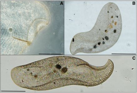
An elusive giant has risen from the muck at the bottom of a Florida canal. It is the ciliate Loxodes rex, a large and shapely thing once thought to be endemic to equatorial Africa. Hunter Hines, a Ph.D. student affiliated with the Harbor Branch Oceanographic Institution, has published the first  record of the species on the North American continent, and has also posted video of the creature on YouTube:

It is not the first time the species has turned up far from its supposed home. Specimens of the big guy have previously been reported from an artificial pond Thailand.  It also turns up on a diversity checklist from São Paulo (though identifications in lists of that kind can’t be independently corroborated). The new study provides better images and metrics, although silver staining and gene sequencing were not done.

Its presence in Florida is interesting, because Loxodes rex is one of the 52 ciliate “flagship species” listed by Wilhelm Foissner as good candidates for testing the hypothesis that some species of ciliate have a restricted geographical range (Foissner, 2008). The ciliates on Foissner’s list are all eye-catching creatures easily seen in the light microscope, difficult to misidentify and unlikely to be overlooked in diversity studies. All of them seem to show some evidence of endemism: that is, they have not been found everywhere in the world, but have turned up only in certain regions. This makes them ideal for proving or disproving the hypothesis that ciliates, like orchids and elephants, have biogeography. As Foissner and his collaborators put it: “[T]he hypothesis of restricted distribution of certain ciliate species must be refused when a considerable number of them is found in all or most biogeographic regions.” (Foissner et al, 2008)

Loxodes rex, from Hines et al, 2016

Loxodes rex, from Hines et al, 2016

To some readers, it might seem odd that there is any controversy about this. Cave snakes and capybaras have biogeography. Why not ciliates? That question takes us back to a hundred-year-old conjecture by two Dutch biologists, Martinus Beijerinck and Baas Becking, concerning the apparent ubiquity of microbial species. Their speculation was that microorganisms, by virtue of their small size and certain special talents (such as the ability to form resting cysts), were not confined by geographical barriers. Every species could exist anywhere in the world, as long as its preferred environmental conditions are available. If an organism is small enough to waft about in the wind, or travel from place to place in the soggy tailfeathers of a bird, its distribution cannot be limited to any particular region. Baas Becking bundled this idea into a phrase so concise and quotable that it has become almost obligatory to repeat it every time the subject of microbial endemism is raised: “Everything is everywhere, but the environment selects.”

This is sometimes dignified as the “Baas Becking hypothesis,” but Foissner argues that is not a proper hypothesis at all, because it is unfalsifiable (Foissner, 2006). After all, if an organism in one pond fails to appear in another, the difference can always be explained by some small dissimilarity in the local conditions. No two ponds provide identical environmental conditions–indeed, there can be an impressive diversity of micro-environments within a single pond, and local conditions can change from one hour to the next. To test the “ubiquity hypothesis” as expressed in Becking’s memorable phrase, you’d need to have two truly identical bodies of water in two distinct biogeographical regions. It is an impractical experiment, because there are simply too many variables.

The main competitor to “everything is everywhere” is the “moderate endemicity model” advanced by Foissner and others, which holds that while as many as 2/3 of microbial species may be cosmopolitan, the rest are found in only certain places. In other words: Some things are not everywhere. This hypothesis is testable, thanks to the terms Foissner himself has set: if a “considerable number” of the flagship species turn out to be cosmopolitan, moderate endemicity is falsified.

By that standard, it does seem that the newly expanded range of Loxodes rex slightly improves the larger case against ciliate endemism–although, it should be said, Hines does not address Foissner’s stipulation that the flagship species must be found in a site that is not “prone to be contaminated by invaders” (Foissner, 2006).  Obviously, the loss of one purported endemic does not constitute a “considerable number,” but the authors of the Loxodes paper also claim to have found “other large ciliates with alleged restricted distributions,” and have already posted video of one, the fairly cool-looking Frontonia vesiculosa. It will be interesting to see what else they turn up.

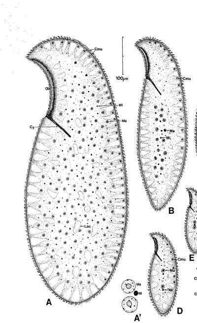
Dragesco's drawing of Loxodes rex (A) next to Loxodes magnus (B). Magnus is big, but rex is the king.

Dragesco’s drawing of Loxodes rex (A) next to Loxodes magnus (B). Magnus is big, but rex is the King.

While Hines et al. do not show stained specimens, the imaging is fairly clear, and the data provided do seem to show that members of the Florida population of Loxodes rex are morphologically close to the ones Jean Dragesco found in Africa. This this can be explained by dispersal, either through natural means (everything is everywhere) or by recent human activity.

Or, it could also be (and what’s a blog for, if not wild speculation?) that both populations are remnants from a single group that split when the Gondwanan parts of the supercontinent Pangaea broke up some 180 million years ago (Florida, unlike most of North America, was largely attached to Gondwana). If that is true, then phylogenetic analysis of the Florida and Africa populations–if anyone ever gets around to it–would show a lot of genetic divergence (a few hundred millions years’ worth), even though morphology of the species has been conserved. Morphological stasis of a species, even over long stretches of geological time, is unexceptional, as Stephen Jay Gould argued at exhausting length in The Structure of Evolutionary Theory, and recent work by Heger et al. on the testate amoeba Hyalosphenia papilio has shown how much genetic diversity can be concealed behind a highly conserved morphology.

If the Florida population of Loxodes rex turns out to be a stable, genetically distinct subgroup, like the cryptic species Hyalosphenia papilio studied by Heger et al., the notion that ciliates have biogeography remains somewhat intact, while the practice of defining species by small numbers of morphological characters suffers another small blow.

Of course, that moves the goal posts for testing the “ubiquity hypothesis,” but I do feel we need to ask whether the ongoing dispute about whether “everything is everywhere” is, in part, an artifact created by simplistic species concepts.

The paper by Hines et al. is Open Access, and can be read here: The First Record for the Americas of Loxodes rex, a Flagship Ciliate with an Alleged Restricted Biogeography.

CLICK for REFERENCES

Feb 182015
 

In my previous post on the lorica-dwelling scuticociliate Calyptotricha pleuronemoides, I mentioned that only two substantial articles have been written about the species since its discovery (apart from brief descriptions in various places). Until yesterday, I’d been unable to find the second article, which appeared in the German microscopy journal Mikrokosmos in 1999. Luckily, one of the co-authors, Martin Kreutz, was kind of enough to send me a copy!

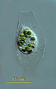
calyptotrichapleuronemoides1_mkw

Calyptotricha pleuronemoides. Image by Martin Kreutz. Source: micro*scope. Click on image for link to source.

Martin tells me he sees the organism frequently in water from the sphagnum ponds of Simmelried, a system of bog lakes, like the one in Ottawa’s Mer Bleue where I sometimes find Calyptotricha. He and Philipp Mayer provide a good redescription of the ciliate, with morphometrics. Their measurements match those of the specimens I’ve found, and agree with those of Phillips and D.S. Kellicott. (All sources give a size range somewhat smaller than that recorded by Alfred Kahl, who gives 50 µm for the length of the cell, and 85 µm for the lorica).

The Mikrokosmos article is difficult to find, so I thought I might give a brief redescription of the species, based on the information collected by Kreutz and Mayer:

Calyptotricha pleuronematoides:  Pleuronematid ciliates, in spindle-shaped hyaline lorica 56-75 µm long, tubular with narrowed openings at either end, 9-13 µm in width. Lorica broadens in the middle to a width of 23-24 µm. Cell body resembling Cyclidium, slightly flattened back to front, 22-35 µm long, 15-24 µm wide, somewhat concave on the ventral surface, where oral apparatus occupies 3/4 of body length. L-shaped undulating membrane, made up of fused cilia, 14 µm long. Caudal cilium 10-12 µm long; roughly 17 somatic kineties, spaced 2-3 µm apart. Most specimens with 8-15 zoochlorellae, colorless examples rare. Single oval macronucleus, with small spherical micronucleus. CV in posterior.

The authors mention that specimens are seldom seen outside of their loricas. Free-swimming individuals move quickly, but not as jerkily as Cyclidium.  I happen to have recorded a free-swimming Calyptotricha, last year, so I might as well post it here:

References:

Kreutz, Martin, and Philipp Mayer. “Artikel-Calyptotricha pleuronemoides-Ein Ciliat in einer Rohre.” Mikrokosmos 88.1 (1999): 27-30.
Feb 132015
 

A few years ago, I looked in a sample of water from a bog lake, and saw something like a hyperactive avocado shifting around inside in a tiny kerosene lamp:

The architect of that pretty dwelling is the ciliate Calyptotricha pleuronemoides. The species and genus were discovered in 1882, in samples from a pond near Hertford, England, by an amateur naturalist named Frederick W. Phillips.  Not much is known about him.  During the 1880s, he was an active member of the Hertfordshire Natural History Society and Field Club, to whom he occasionally read essays on “The Protozoa of Hertfordshire,” based largely on the classification scheme in William Saville Kent’s Manual of the Infusoria.  He was a Fellow of the Linnean Society of London, and he found and named a few new taxa.

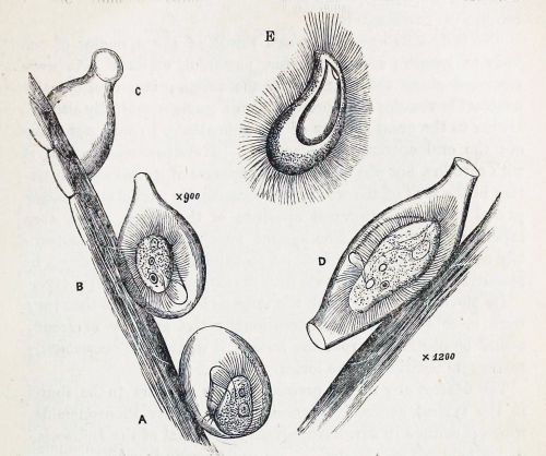
In his very first glimpse of the creature, Phillips was lucky enough to catch it in the act of building its lorica. “At first sight,” he writes, “I thought it was an embryonic or encysted stage of some monad; but upon applying a magnifying power of some 900 diameters, I observed that it possessed a singular vibratile membrane, closely resembling that which characterizes the members of the family Pleuronemidae.” A week later, Phillips looked at it again, and discovered that “the lorica had increased in size, and that one end was elongated into a teat-like form.” At this stage, he accidentally allowed the sample to dry out, leaving the organism’s empty, half-finished lorica still attached to a strand of pond-weed. He made a nice drawing of what he’d seen.

Calyptotricha pleuronemoides from Phillips resized

A. First stage B. The same, further developed C. End view of lorica D. The perfect animal E. Ventral view (adapted from Phillips)

To modern readers, accustomed to the impersonal, passive style of scientific writing–“samples were collected,” “living cells were isolated and observed”–there is something pleasingly candid about the way Victorian naturalists report their findings. Phillips doesn’t just describe his new genus, he spins us the tale of its discovery, including the mishap that destroyed his first specimen, and his initial misreading of the oval shell, after which he takes us to the very moment of discovery when he exposed the creature’s true nature by “applying a magnification of 900 diameters.” Something about that reminds me of the exploration literature of the same period. It’s probably not just an accident of style: Victorian microscopists were explorers. Superior lenses and stains had opened up a miniature Dark Continent on their laboratory benches, and a gentleman adventurer from somewhere like Hertfordshire could now penetrate these hidden realms, returning with breathless accounts of what he had seen. A session at the microscope was an expedition into the unknown.

In our time, researchers are expected to pile up some data before going to print, and nobody would attempt to erect a new ciliate genus on the basis of a brief observation of a few specimens. No doubt that is a good thing: the 19th century left a big legacy of poorly defined taxa, many of which are still desperately in need of revision.  But this kind of field work, as sketchy and dilettantish as it might seem now, has largely been put to one side without really being replaced by anything better.  Outside of a few centers of activity, ciliate field work has slowed to a crawl.  Consider the fact that 132 years after Phillips wrote his three-page note on Calyptotricha pleuronemoides it is still one of only two substantial treatments of the species, and the only source that describes the construction of its curious lorica.  Anyone who wants to know more about this ciliate than its name, has to travel back to the 19th century.

poke bonnet

A straw poke-bonnet, from the early 19th century. (Click for source)

Needless to say, the old information is not always reliable.

Phillips perceived immediately, and rightly, that Calyptotricha is closely related to the more common ciliate Pleuronema. Like its cousin, it is equipped with a large, billowing membrane that runs along the right side of its oral aperture. However, Phillips badly misunderstood the shape of this structure, describing it as “a membranous trap, or velum, which in form resembled the old-fashioned poke-bonnet.”

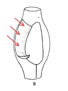
When I first read that passage, the comparison to a “poke-bonnet” confused me. The undulating membrane of pleuronematid ciliates is shaped something like a sail, or a flag: a sheet of fused cilia running along one side of the organism’s mouth. Phillips, however, interpreted this structure (which, admittedly, is very difficult to see clearly in the light microscope) as a sort of hood or canopy covering the oral aperture of the ciliate. If you look closely at his illustration, you can see that he has drawn it as a baggy tube.

Calyptotricha's undulating membrane resembles a sail or banner (image adapted from Colin R. Curds, British and Other Freshwater Ciliated Protozoa)

The true shape of Calyptotricha’s undulating membrane (image from Colin R. Curds, British and Other Freshwater Ciliated Protozoa, with arrows added)

Evidently, it was this imagined resemblance to a poke-bonnet that prompted him to give the genus its curious name, Calyptotricha, constructed from the Greek calyptos (“veiled” or “covered”) and trich (“hair”). It seems the “haired” holotrichous ciliate reminded him of a woman’s head, on top which the membrane sits like an old-fashioned hat!

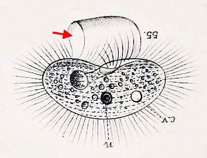
It’s an example of how expectation shapes observation. In interpreting this membrane as an enclosed hood, he was deferring to an earlier error by his illustrious contemporary William Saville Kent. Writing about Pleuronema, Kent says: “[T]his membranous trap may be appropriately compared with the extensile hood of a carriage or an outside windowshade forming, when expanded, a capacious hood-shaped awning, and when not in use being packed away in neat folds close around the animalcule’s mouth.”

The “extensile hood” Kent mentions was a common convenience on carriages of his day, and provided a compelling mechanical analogy for the “neat folds” with which he imagined Pleuronema pulled back its velum.

Barouche image 2Here, for comparison, is Kent’s illustration of Pleuronema chrysalis, which I’ve inverted to showcase its “extensile hood.”

Pleuronema chrysalis, from W. S. Kent's A Manual of Infusoria. Put wheels on it, and you have a chuck wagon.

With wheels, it would make a good chuck wagon.

To modern workers familiar with the morphology of hymenostome ciliates, as revealed in specimens that have been stained with silver, this is an implausible design. However, to Kent, who had done pioneering work on choanoflagellates, it seemed reasonable to speculate that Pleuronema’s hood might share “a distant homological relationship” with the “delicate funnel-shaped membranes” found in the collared flagellates, which really do wear something a bit like a straw poke-bonnet (but on the back end of the cell).

Finally, since we’ve been talking about Pleuronema and her sisters, I’ll post some footage of one, quietly browsing on bacteria in water taken from a tidal pool on the coast of Maine:

REFERENCES

Oct 092014
 

As I might have mentioned already, my favorite protists are the shaggy, shapely, fast-moving ciliates. They have a lot to offer the idle protist-ogler. As a group, they include some of the largest and most ridiculous-looking microbes in the pond. Many are easy to identify without expensive equipment or special techniques. Some, like the noodle-necked Lacrymaria olor, can be recognized at a glance in the light microscope, even at low magnification. Others, like the stately Stentors, may need closer inspection for a species-level classification, but can still be identified by prominent features such as the colour of the cell, or the shape and distribution of various organelles.

Unfortunately, not all ciliates are so easy to tell apart.  Some are like the “little brown birds” that plague neophyte birders, and can only be distinguished from one another by very close observation under exacting conditions.  And many, I’m sorry to say, are pretty much impossible to identify, even to genus level, without the help of special stains that expose distinctive patterns in the cilia on the surface of the cell body.

The classic technique for exposing these structures is to fix the cells in some noxious and foul-smelling substance and then soak them in solutions containing various compounds of silver. Certain parts of the organism–most conveniently, for our purposes, the ciliary rows and the nuclei–are “argentophilic,” which is to say they stain darkly when exposed to silver. The ability to selectively stain these organelles revolutionized ciliate taxonomy in the second half of the 20th century, and it is still the most important technique available to modern ciliatology.

Despite my particular interest in ciliates, I’d never tried it until just a few days ago.

I’ve been slow to get around to this, mainly because it’s hard to do. Even the easiest methods of silver staining call for a cupboard full of powders and solvents, none of which is available at Shopper’s Drug Mart, and some of which must be handled and stored very thoughtfully. To procure the ingredients I had to find suppliers willing to do business with an individual buyer, and in some cases I had to pay special transport fees.

lab reagents

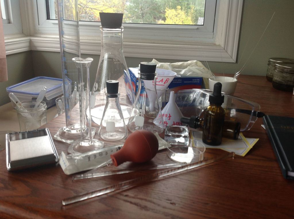
Then, of course, I had to assemble the equipment required to use this stuff safely: graduated cylinders, flasks, funnels, fixing jars, an accurate scale, syringes, latex gloves, etc.

lab stuff

And finally, I had to acquire a bunch of new skills.  I haven’t stood at a lab bench since the ninth grade (40 years ago, if you can believe it), so I had to learn how to do simple tasks, like weighing, pouring and mixing.  Fortunately, before undertaking any of this, I had the foresight to culture a full-sized biochemist, which was quite expensive and took about 23 years.  He is currently living in my basement, and was very helpful at several points.

Here, then, is my first attempt at staining a plain old Paramecium by one of the silver carbonate methods:

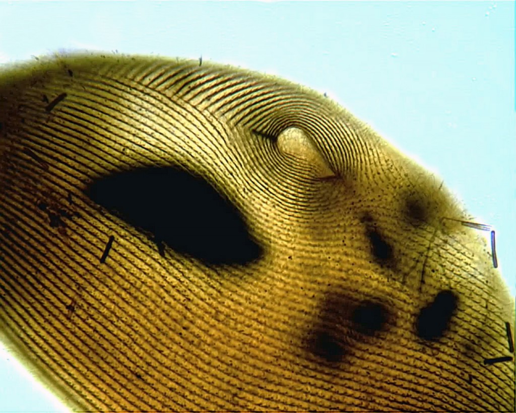
Silver carbonate Paramecium

Yes, there’s a lot of room for improvement, but, frankly, I’m delighted that it worked at all.

Here’s one more from the same slide, a specimen of a common hymenostome ciliate with the curious name of Glaucoma:

?????????????

The impregnation could be more uniform, the focus could be sharper and the hot spot from the microscope lamp is downright annoying. However, I can count the kineties and easily see the shape of the macronucleus.  That’s a step forward, for me.

The protocol I followed is the one developed by Augustin, Foissner and Adam in 1984 (described in Foissner’s updated guide to basic methods for ciliate taxonomy). I’m told that the original Fernandez-Galiano method gives more consistent results, so I’ll try that next.


Dec 182013
 

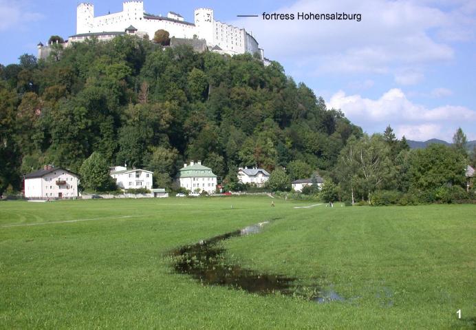
Early last year, the mayor of Salzburg proudly announced the creation of a new conservation zone around this “globally unique ‘natural monument'”:

Krauthugel

Click image for source,

The newly protected feature is not that rocky bluff, Festungsberg hill, or the 11th-century fortress that sits on top of it. It is the long, narrow puddle in the foreground.  This is Krauthügel Pond, an ephemeral body of water barely 30 cm deep where researchers have found 121 species of ciliates, ten of them previously undescribed. Because of these organisms–five of which have not been found elsewhere–Salzburg now possesses the world’s first second “Natural Monument for Single-celled Organisms.” A protist wildlife sanctuary!

The pond comes and goes during the year, appearing after heavy rains on a raised agricultural plot known as Krauthügel, or “cabbage hill.” The bed in which it lies is thought to be the remains of an old stream, whose natural course might have been altered by agriculture during the middle ages. Since then, roadwork and urban development have isolated the body from other surrounding channels.

From 1789 until 1960, the field was used for raising vegetables.  After that, it became a pasture. For about thirty years, cows trampled the soft turf and nourished the local microbes with their  manure, creating what ecologists call a “eutrophied pond.” In other words, a cattle wallow, or slough.

This is not what you could call a pristine natural environment. It doesn’t shelter any large, charismatic animals. It is not particularly scenic, when it can be seen at all (much of the year, the “pond” is dry). In short: it’s hard to imagine a patch of ground less likely to be singled out for conservation.

However, Krauthügel Pond has something your local ditches and mudholes lack: proximity to Wilhelm Foissner, an astonishingly productive ciliatologist who happens to live and work in Salzburg.

Foissner

Wilhelm Foissner (Click Image for Source)

Arguably, the “natural wonder” here is not so much Krauthügel pond as Professor Foissner, whose vast body of work looms over modern ciliate systematics like the Festungsberg itself. With five or six hundred publications to his name–at least three hundred in peer-reviewed journals–Foissner, working alone or in collaboration, has discovered and described over 500 new protist species.  If there were new ciliates in your cow field, he would be the man to find them.

Actually, to see a new species is not that unusual.  Likely, we all run across undescribed organisms, from time to time, without knowing it. The little red bug that alights on your arm might be something never before recorded in the literature, if only you knew. Place samples of local mud under the microscope, and you are quite likely to find organisms that don’t yet have names. Of course, it’s one thing to see something new as it paddles by, and quite another to know what you have seen. To properly document your discovery, and publish the news of it, requires skills and technology that are in extremely short supply.

So, these ciliates were pretty lucky to have been born in Salzburg, near one of the few people in the world with the ability (and inclination) to see them for what they are and lobby for their protection. It raises some interesting questions.

First, how exceptional is the microbe diversity that has been preserved in Krauthügel?  In a report on the the pond, published earlier this year, Fenton P.D. Cotterill and his co-authors compare the ciliate species count at their location to various “well-investigated ephemeral waters” in other parts of the world: two meltwater ponds in Southwestern Ontario, a roadside puddle in Namibia, a rock pool in Venezuela, a meadow in Hungary, and several other choice spots.  They find that the Salzburg pond is in the “upper range” for total number of species, but only in the “middle range” for the number of new species.

Evidently, the old cabbage field supports a high–but far from unique–diversity, and when closely probed by the best protistologists in the business, it yields about the expected number of new organisms. A rich but fairly ordinary body of water, it seems.  Why single it out for protection?

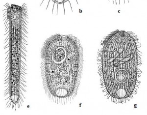
Three species only found at Krauthügel  e)  Semispathidium pulchrum f) Papillorhabdos multinucleatus g) Fuscheria nodosa salisburgensis

Three species only found at Krauthügel e) Semispathidium pulchrum f) Papillorhabdos multinucleatus g) Fuscheria nodosa salisburgensis

There are a couple of reasons. First, as the authors point out, appeals for conservation are usually based “on the narrow distribution of one or several species and their habitat, or of species and habitats endangered by human activities.”  If a forest supports the only known population of Sibree’s Dwarf Lemur, we have reason to preserve it, because if we don’t, we can expect to lose that species forever. By that standard–provided we suppress the size-bias that can make us indifferent to the fate of a microbial species–the case for protecting Krauthügel is pretty strong.  As of April, 2013, five of the the ten new species found there had “not been reported from any other locality.”  Until they turn up elsewhere, those five species are assumed to be “endemic” to Salzburg (that is, restricted to that area).  Given the scarcity of competent ciliatologists in other parts of the world, they may remain so for quite a while.

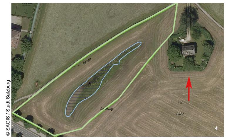
Whether they turn out to be truly endemic or not, it is indisputable that the organisms in the pond were “endangered by human activities”. In 2010, as part of an art project, somebody filled it in with earth. Imagine the alarm of researchers who had been studying the site for decades when they found out their protists had been buried! It was this event that prompted investigators to call for protection, resulting in the restoration of the pond to its previous condition and the creation of a buffer zone around it:

Buffer zone around Krauthügel Pond

Protected zone around Krauthügel Pond

And that brings us to the second reason for conserving this puddle: thanks to the work that had already been done there, it has become the “type locality” for some eighteen species (eight new species, and ten redescribed taxa). The significance of this might require a bit of explanation.

amblyodus taurus

Amblyodus taurus (click image for source)

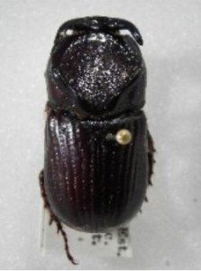
When a biologist names a new taxon, the usual practise is to select a particular fixed specimen, or group of specimens, as the “type,” and (ideally) to deposit that specimen in a permanent collection somewhere, available to other researchers.  This provides a permanent concrete reference, so there can be no ambiguity about what we really mean when we say Utricularia floridana (a species of carnivorous plant), or Amblyodus (a genus of beetle).  If need be, we can point to a certain bug on skewered on a certain pin and say, “There! Amblyodus means that.”

The site at which the type specimen was collected becomes the “type locality,” where one might expect to find others of the same breed.  That locality is especially important to protist taxonomists. Protists are small and fragile, and fixed type specimens of older named organisms are rarely available.  Even when permanent slides exist, they can be lost, or simply deteriorate over time.  If we know the type location where our guys were originally found, we can go look for them there. In theory. But if the place at which the work was done has been drained or paved, and no type material exists, the identities of the species found there can be lost in taxonomic noise.

What is being conserved at Krauthügel is, at least in part, the scholarly work that has already been done there.  It is a body of acquired biological knowledge, and not just the organisms themselves, that is being protected.  From this point of view, environmental conservation can be similar to task that museum and art conservators do, preserving the best products of human effort for future generations.

Where does that leave all the ponds that haven’t been, and likely never will be, studied?  In spring, when I drive through the countryside where I live, I see ephemeral pools by the hundreds and thousands. They flash by in the car window, mile after mile: beaver ponds, ditches, mill pools, flood plains, and wide shallow puddles in fields where cows dip their muzzles and drop their nutritious poops. Some will have less protist diversity than Krauthügel, a few may have more, but none will ever enjoy the benign oversight of Wilhelm Foissner.

But what if more research were being done on these bodies of water–a protistologist for every puddle!–and more ponds found worthy of conservation? It is not clear where that road goes. If the Krauthügel initiative stirred up any controversy in Salzburg, there’s no record of it in the article, or the press release, but it’s not hard to anticipate the kind of pushback we’d see if similar initiatives were tried here.  Attempts to control the use of private land arouse deep and incredibly long-lived resentments.  Twenty-five years after efforts to conserve habitat for the Northern Spotted Owl in the PNW, anti-environmentalists are still seething and sneering. In some circles, the words “spotted owl” have become a kind of shorthand for “meddlesome tree-hugging morons who place a higher value on a stupid bird than the lives and livelihoods of hardworking humans.” Imagine the volcano of outrage that might erupt over the mandated protection of a one-celled organism! We would never hear the end of it.

All the same, the idea of protecting protist habitat has a lot of appeal for me.  Down the road from my house in the Gatineau hills, there’s a group of ephemeral ponds where I like to gather samples. Within a few years, they will almost certainly be filled in, as the land is subdivided for new housing. I’ve watched those ponds for several seasons, and hate the idea of losing their amazing microscopic diversity. If, as is statistically probable, they contain a few new species, there might even be grounds for conservation. However, it is pretty certain that the bulldozers will get to any new organisms before I gain the competence and resources to find and describe them.

Still,  I find it a little comforting to remind myself of the very different scale and speed of life at the microscopic level. When you are a hundred microns long, from tip to tail, a puddle is a lake, and a pond is an ocean. An hour is a year!

In a few days, a rain-filled tire rut can burst into startling diversity, like a miniature coral reef. Species bloom in quick succession, replacing and displacing one another. Each one changes the chemistry, light-permeability and nutrient load of the water, conditioning the environment to suit certain organisms, all of whom, in turn, will alter the water around them. Accidents of geography (a floating leaf,  a ball of dung) make opportunities for some organisms, and extinguish all hope for others. Things progress quickly. If you return to the tire-rut every day and follow its progress with the help of a microscope, it can seem like looking at a time-lapse film. In the “big” world, environments change in much the same way, but over longer periods of time: forests encroach on prairies, then recede; wetlands take shape, silt up and vanish; animals come and go.  At the microscopic level, shifts in populations may happen in hours, instead of years, and change is unremitting. “Ecological balance” is never achieved: things happen, then more things happen. Some species flourish, others fade from view. Then one day the hot sun dries it all up, and the little creatures climb back into their resting cysts, as their habitat reverts to grass.

———————————
References:

Cotterill, Fenton PD, et al. “Conservation of Protists: The Krauthügel Pond in Austria.” Diversity 5.2 (2013): 374-392.