In my previous post on the lorica-dwelling scuticociliate Calyptotricha pleuronemoides, I mentioned that only two substantial articles have been written about the species since its discovery (apart from brief descriptions in various places). Until yesterday, I’d been unable to find the second article, which appeared in the German microscopy journal Mikrokosmos in 1999. Luckily, one of the co-authors, Martin Kreutz, was kind of enough to send me a copy!
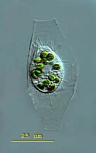
Calyptotricha pleuronemoides. Image by Martin Kreutz. Source: micro*scope. Click on image for link to source.
Martin tells me he sees the organism frequently in water from the sphagnum ponds of Simmelried, a system of bog lakes, like the one in Ottawa’s Mer Bleue where I sometimes find Calyptotricha. He and Philipp Mayer provide a good redescription of the ciliate, with morphometrics. Their measurements match those of the specimens I’ve found, and agree with those of Phillips and D.S. Kellicott. (All sources give a size range somewhat smaller than that recorded by Alfred Kahl, who gives 50 µm for the length of the cell, and 85 µm for the lorica).
The Mikrokosmos article is difficult to find, so I thought I might give a brief redescription of the species, based on the information collected by Kreutz and Mayer:
Calyptotricha pleuronematoides: Pleuronematid ciliates, in spindle-shaped hyaline lorica 56-75 µm long, tubular with narrowed openings at either end, 9-13 µm in width. Lorica broadens in the middle to a width of 23-24 µm. Cell body resembling Cyclidium, slightly flattened back to front, 22-35 µm long, 15-24 µm wide, somewhat concave on the ventral surface, where oral apparatus occupies 3/4 of body length. L-shaped undulating membrane, made up of fused cilia, 14 µm long. Caudal cilium 10-12 µm long; roughly 17 somatic kineties, spaced 2-3 µm apart. Most specimens with 8-15 zoochlorellae, colorless examples rare. Single oval macronucleus, with small spherical micronucleus. CV in posterior.
The authors mention that specimens are seldom seen outside of their loricas. Free-swimming individuals move quickly, but not as jerkily as Cyclidium. I happen to have recorded a free-swimming Calyptotricha, last year, so I might as well post it here:
References:
Kreutz, Martin, and Philipp Mayer. “Artikel-Calyptotricha pleuronemoides-Ein Ciliat in einer Rohre.” Mikrokosmos 88.1 (1999): 27-30.
A few years ago, I looked in a sample of water from a bog lake, and saw something like a hyperactive avocado shifting around inside in a tiny kerosene lamp:
The architect of that pretty dwelling is the ciliate Calyptotricha pleuronemoides. The species and genus were discovered in 1882, in samples from a pond near Hertford, England, by an amateur naturalist named Frederick W. Phillips. Not much is known about him. During the 1880s, he was an active member of the Hertfordshire Natural History Society and Field Club, to whom he occasionally read essays on “The Protozoa of Hertfordshire,” based largely on the classification scheme in William Saville Kent’s Manual of the Infusoria. He was a Fellow of the Linnean Society of London, and he found and named a few new taxa.
In his very first glimpse of the creature, Phillips was lucky enough to catch it in the act of building its lorica. “At first sight,” he writes, “I thought it was an embryonic or encysted stage of some monad; but upon applying a magnifying power of some 900 diameters, I observed that it possessed a singular vibratile membrane, closely resembling that which characterizes the members of the family Pleuronemidae.” A week later, Phillips looked at it again, and discovered that “the lorica had increased in size, and that one end was elongated into a teat-like form.” At this stage, he accidentally allowed the sample to dry out, leaving the organism’s empty, half-finished lorica still attached to a strand of pond-weed. He made a nice drawing of what he’d seen.
A. First stage B. The same, further developed C. End view of lorica D. The perfect animal E. Ventral view (adapted from Phillips)
To modern readers, accustomed to the impersonal, passive style of scientific writing–“samples were collected,” “living cells were isolated and observed”–there is something pleasingly candid about the way Victorian naturalists report their findings. Phillips doesn’t just describe his new genus, he spins us the tale of its discovery, including the mishap that destroyed his first specimen, and his initial misreading of the oval shell, after which he takes us to the very moment of discovery when he exposed the creature’s true nature by “applying a magnification of 900 diameters.” Something about that reminds me of the exploration literature of the same period. It’s probably not just an accident of style: Victorian microscopists were explorers. Superior lenses and stains had opened up a miniature Dark Continent on their laboratory benches, and a gentleman adventurer from somewhere like Hertfordshire could now penetrate these hidden realms, returning with breathless accounts of what he had seen. A session at the microscope was an expedition into the unknown.
In our time, researchers are expected to pile up some data before going to print, and nobody would attempt to erect a new ciliate genus on the basis of a brief observation of a few specimens. No doubt that is a good thing: the 19th century left a big legacy of poorly defined taxa, many of which are still desperately in need of revision. But this kind of field work, as sketchy and dilettantish as it might seem now, has largely been put to one side without really being replaced by anything better. Outside of a few centers of activity, ciliate field work has slowed to a crawl. Consider the fact that 132 years after Phillips wrote his three-page note on Calyptotricha pleuronemoides it is still one of only two substantial treatments of the species, and the only source that describes the construction of its curious lorica. Anyone who wants to know more about this ciliate than its name, has to travel back to the 19th century.
A straw poke-bonnet, from the early 19th century. (Click for source)
Needless to say, the old information is not always reliable.
Phillips perceived immediately, and rightly, that Calyptotricha is closely related to the more common ciliate Pleuronema. Like its cousin, it is equipped with a large, billowing membrane that runs along the right side of its oral aperture. However, Phillips badly misunderstood the shape of this structure, describing it as “a membranous trap, or velum, which in form resembled the old-fashioned poke-bonnet.”
When I first read that passage, the comparison to a “poke-bonnet” confused me. The undulating membrane of pleuronematid ciliates is shaped something like a sail, or a flag: a sheet of fused cilia running along one side of the organism’s mouth. Phillips, however, interpreted this structure (which, admittedly, is very difficult to see clearly in the light microscope) as a sort of hood or canopy covering the oral aperture of the ciliate. If you look closely at his illustration, you can see that he has drawn it as a baggy tube.
The true shape of Calyptotricha’s undulating membrane (image from Colin R. Curds, British and Other Freshwater Ciliated Protozoa, with arrows added)
Evidently, it was this imagined resemblance to a poke-bonnet that prompted him to give the genus its curious name, Calyptotricha, constructed from the Greek calyptos (“veiled” or “covered”) and trich (“hair”). It seems the “haired” holotrichous ciliate reminded him of a woman’s head, on top which the membrane sits like an old-fashioned hat!
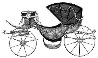
It’s an example of how expectation shapes observation. In interpreting this membrane as an enclosed hood, he was deferring to an earlier error by his illustrious contemporary William Saville Kent. Writing about Pleuronema, Kent says: “[T]his membranous trap may be appropriately compared with the extensile hood of a carriage or an outside windowshade forming, when expanded, a capacious hood-shaped awning, and when not in use being packed away in neat folds close around the animalcule’s mouth.”
The “extensile hood” Kent mentions was a common convenience on carriages of his day, and provided a compelling mechanical analogy for the “neat folds” with which he imagined Pleuronema pulled back its velum.
Here, for comparison, is Kent’s illustration of Pleuronema chrysalis, which I’ve inverted to showcase its “extensile hood.”
Finally, since we’ve been talking about Pleuronema and her sisters, I’ll post some footage of one, quietly browsing on bacteria in water taken from a tidal pool on the coast of Maine: