Nov 182015
 

I’ve finally identified a thing I found in my mouth nearly 25 years ago. Unfortunately, solving that old puzzle has left me with a new one concerning a protozoan normally found in the hindgut of cockroaches and termites.

I ran across it in the fall of 1990. My wife was a first-year medical student and had borrowed a microscope to study samples of human tissue for a course in histology. In those days, I was more interested in poetry than protozoa, but I was even more interested in procrastination. So, one morning I put my doctoral thesis aside and took the loaner ‘scope for a spin.

Her prepared histology slides–fixed cells stained in lurid pinks and powder blues–did not hold my attention for long. I wanted to see something alive and wriggling. I’d read about dust mites, so I put some floor-fluff on a slide, but that was disappointing: nothing but hair, skin flakes and coloured cloth fibres. Then, I sampled some water from a soup pot that had sat in the sink overnight, which was a little better. There were bacteria there, thousands of them, jostling and jiggling in the milky broth. But bacteria are boring.

Mouth thing

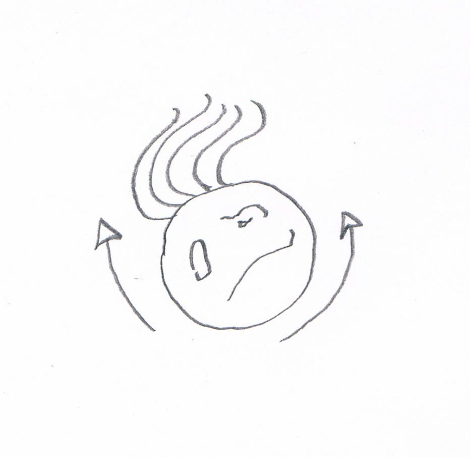
A reconstruction of The Thing. (The arrows are supposed to show how it moved. Yes, it looks like the cell has arms, but I don’t feel like drawing it again).

Finally, I swabbed some saliva from the back of my mouth and that was more like it. If you’ve ever done this, you’ll know what I found: big, ragged oral epithelial cells, swirling rafts of intriguing organic stuff, plump leukocytes that had recently eaten a bacterial meal. Then something truly bizarre floated into view. It was round and mottled, like a degenerate happy face with a crop of lank hairs sprouting from one side. It was rotating back and forth with a hypnotic, rocking motion: clockwise, then counterclockwise, then clockwise again. With each change of direction, the strands moved together in a languid arc.

Amazing! A parasite was living in my mouth, and it had hair like a rock star. What could it be?  I grabbed a bus down to the McGill Life Sciences Library and pulled some parasitology texts from the stacks. From these, I determined that this creature had to be some kind of “flagellate” (the word was new to me). I leafed through the mug shots, blurred micrographs of Trichomonas, Chilomastix, Giardia, Retortamonas and all their troublesome colleagues, but I couldn’t find my guy anywhere. The known flagellate parasites were all were too pinched, or too pointy, and none had the lush head of hair I’d seen on my creature.

A couple of nights later, a burglar broke into our place and stole the microscope, which put an end to my amateur parasitology for a couple of decades. I went back to my real work, but never forgot that my mouth had miniature glam rockers living inside it. Years later, when I returned to microscopy (taking procrastination to a whole new level), the first thing I did was try to find those flagellates again. I swabbed my palate, scraped my tongue and spat on slide after slide, without success. By then, I had learned a lot more about protists, and I had acquired a good library of field guides and protozoology texts, none of which recorded anything quite like the organism I’d seen. I began to doubt my memory.

Then, just two days ago, a friend from Uruguay sent me a link to a video from Mexico, showing a presumed protozoan found in the bronchial lavage of a patient who had nearly drowned in a river. And there it was again, the Thing from my Mouth!

That rocking motion is exactly what I had seen in 1990. The video was posted to YouTube as an unknown “ciliado” (ciliate), and superficially it does resemble one. At first, it struck me as an overripe oligotrich, possibly bloated from osmotic pressure and moving poorly. But the way it was moving looked all wrong, even for an unhealthy specimen. That repetitive, ineffectual rocking movement was not like the familiar motion of the fused cilia (adoral polykinetids) that surround the cellular mouth of oligotrich ciliates, or any of the ciliary wreaths and fringes I knew from other free-living ciliophora. The rocking suggested something sessile that had come loose from its substrate, which reminded me that this “ciliate” had been found in bronchealveolar lavage fluid (BALF).  As it happens, we all have ciliated cells lining some of our nasal and bronchial cavities, which perform the useful task of clearing mucus and debris from the lungs. The ciliated cells stay fixed in one place, and waste matter sort of “crowd surfs” over them, passing from one cell to the next on its way out the door. Could this be a detached bronchial cell?

A search within YouTube turned up another video, this one from the Diagnostic Bacteriology Laboratory at Singapore General Hospital. It shows a large clump of bronchial epithelium with the cilia busily working:

In this sample, the cells are all fixed in place and firmly attached to one another, so we don’t see the feeble rocking movement of the free-floating cell. Still, I’m persuaded me that the cells in both videos, as well as the one I’d seen back in 1990, are all ciliated nasal or bronchial cells.

And this brings me to the new puzzle. A comment on the second video sent me to an intriguing post at a pathology blog called Microcosm, where I learned something fascinating and, I’m sorry to say, a bit revolting. It seems there have been medical case reports of humans being infected by a lushly flagellated protozoan called Lophomonas blattarum, normally found living commensally in the intestines of cockroaches or termites.

lophomonas blattarum from Kent

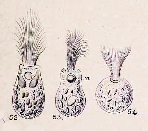
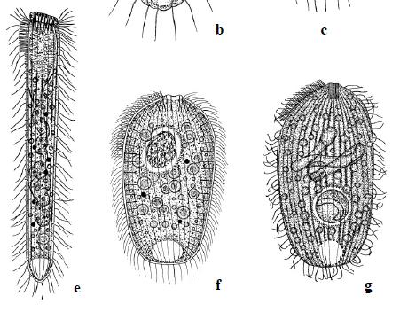
Lophomonas blattarum, from William Saville Kent, A Manual of Infusoria, 1881.

L. blattarum has been known since 1860, when it was found and described by the great Samuel Friedrich Stein. Reports of respiratory infections in humans are more recent, dating back only as far as 1993 when the first case was reported in the Chinese Journal of Parasitology and Parasitic Diseases. Since then, many dozens of similar cases have been reported, which has lead to a growing belief, among some researchers, that Lophomonas blattarum constitutes “a potentially important cause of bronchopulmonary infection and respiratory symptoms.” (Martinez-Girón and van Woerden, 2013a).

The problem is, it might not be a genuine infection at all. In all of the published case reports of Lophomonas infection, the organisms were identified by light microscopy, either from living samples or stained preparations. To date, there have been no molecular studies of the purported parasite. Identifications have been based entirely on morphological features and symptomology.

One ultrastructural study has been done, but it does not support the theory that lophomoniasis is a genuine infective disease. The 12-author paper was published in Chinese, but from the English abstract (and by laboriously feeding the text to Google Translate one paragraph at a time), I gather that the researchers used optical and electron microscopy to examine mobile cells from BALF of 6 patients, which they found to be morphologically unlike Lophomonas. They also reviewed the existing literature on Chinese cases of lophomoniasis, and reached the following conclusion: “In the past 20 years, all the diagnosed cases as pulmonary Lophomonas blattarum infection reported in our country were misdiagnosed. Currently, there is no evidence to show Lophomonas blattarum as a pathogen resulting in pulmonary infection.” (Mu XL, et al., 2013)  According to this study, the Lophomonas infections recorded in the literature were actually misidentified bronchial cells.

Lophomonas blattarum from He et al

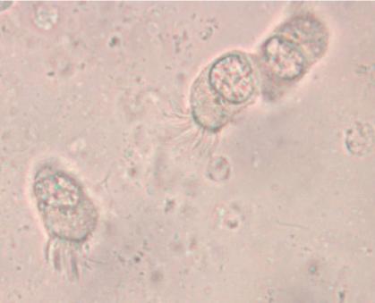
A typically murky micrograph of cells identified as Lophomonas blattarum in He et al, 2011. Martinez-Girón and van Woerden consider these to be misidentified bronchial epithelial cells.

Other workers, while agreeing that confusion between epithelial cells and Lophomonas has occurred, continue to maintain that many cases of infection are genuine. Martínez-Girón and van Woerden, who have published more about lophomoniasis than anyone, have responded to their Chinese colleagues by reaffirming their opinion that “the observation under light microscopy of this multiflagellated protozoon in symptomatic patients, who respond positively to antiprotozoal therapy, can reasonably be described as bronchopulmonary lophomoniasis.” (Martínez-Girón and van Woerden, 2013b)


Lophomonas blattarum image resized

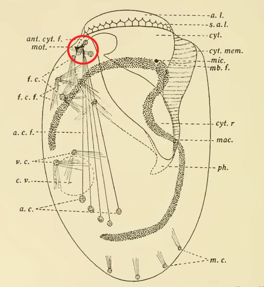
The fine structure of Lophomonas blattarum (Beams et al, 1961, adapted from Kudo) . Note the wine-glass shaped calyx (CY), axial filament (AXF) and parabasal “collar” of tubules (PNT), all visible in the light microscope.

As someone who is usually more interested in the taxonomy of organisms than the pathologies they cause, I’m struck by the emphasis clinicians place on disease and symptoms rather than the creature itself. From a clinical point of view, the case for infection is strong: a given patient has respiratory symptoms, some apparently-flagellated cells are observed in bronchial lavage fluid, and the condition clears up with treatment by metronidazole, which is, after all, the preferred treatment for another metomonad parasite, Trichomonas vaginalis. These are all important facts that are hard to dismiss if you approach the subject from a clinical point of view. However, from a purely protistological perspective, we might note that identification by light microscopy is often unreliable, especially with such small cells; the micrographs in the published literature are usually quite poor, and may depict cells in an unnatural condition; the morphological characters used in these case studies tend to be weak, while stronger diagnostic characters, like the fine structures shown in the diagram to the right, are not mentioned; and, finally, the organism in question is a deep-branching anaerobe adapted to life in the intestines of insects of the order Blattodea, and it is a little suprising for it to flourish in a human lung. An organism’s habitat is a strong clue to its identity; this one is normally found in the airless hindgut of certain bugs, and possibly in the poop of certain bug-eating birds. Given that these flagellates, if that is what they are, were discovered in an untypical environment, a careful taxonomist might wait for more detailed morphological information, or, better yet, molecular data, before settling on a species-level identification. Where are the calyx and axial filament, which Richard Kudo managed to see in 1926? Where are the tubules of the parabasal body, which should encircle to the top of the cell like the ruffled collar of an Elizabethan gentleman?  From what I’ve seen, they’re barely mentioned in the literature on lophomoniasis.

So we have a strong clinical profile combined with unimpressive taxonomic evidence, leaving us with a question that can’t be resolved until somebody succeeds in sequencing these cells. As Martinez-Girón and van Woerden conclude, “The development of a technique to culture the organism or the use of molecular techniques is required to resolve the issue.” If anybody is working on that, it will be interesting to see the results.

——————

REFERENCES (CLICK HERE)


Feb 132015
 

A few years ago, I looked in a sample of water from a bog lake, and saw something like a hyperactive avocado shifting around inside in a tiny kerosene lamp:

The architect of that pretty dwelling is the ciliate Calyptotricha pleuronemoides. The species and genus were discovered in 1882, in samples from a pond near Hertford, England, by an amateur naturalist named Frederick W. Phillips.  Not much is known about him.  During the 1880s, he was an active member of the Hertfordshire Natural History Society and Field Club, to whom he occasionally read essays on “The Protozoa of Hertfordshire,” based largely on the classification scheme in William Saville Kent’s Manual of the Infusoria.  He was a Fellow of the Linnean Society of London, and he found and named a few new taxa.

In his very first glimpse of the creature, Phillips was lucky enough to catch it in the act of building its lorica. “At first sight,” he writes, “I thought it was an embryonic or encysted stage of some monad; but upon applying a magnifying power of some 900 diameters, I observed that it possessed a singular vibratile membrane, closely resembling that which characterizes the members of the family Pleuronemidae.” A week later, Phillips looked at it again, and discovered that “the lorica had increased in size, and that one end was elongated into a teat-like form.” At this stage, he accidentally allowed the sample to dry out, leaving the organism’s empty, half-finished lorica still attached to a strand of pond-weed. He made a nice drawing of what he’d seen.

Calyptotricha pleuronemoides from Phillips resized

A. First stage B. The same, further developed C. End view of lorica D. The perfect animal E. Ventral view (adapted from Phillips)

To modern readers, accustomed to the impersonal, passive style of scientific writing–“samples were collected,” “living cells were isolated and observed”–there is something pleasingly candid about the way Victorian naturalists report their findings. Phillips doesn’t just describe his new genus, he spins us the tale of its discovery, including the mishap that destroyed his first specimen, and his initial misreading of the oval shell, after which he takes us to the very moment of discovery when he exposed the creature’s true nature by “applying a magnification of 900 diameters.” Something about that reminds me of the exploration literature of the same period. It’s probably not just an accident of style: Victorian microscopists were explorers. Superior lenses and stains had opened up a miniature Dark Continent on their laboratory benches, and a gentleman adventurer from somewhere like Hertfordshire could now penetrate these hidden realms, returning with breathless accounts of what he had seen. A session at the microscope was an expedition into the unknown.

In our time, researchers are expected to pile up some data before going to print, and nobody would attempt to erect a new ciliate genus on the basis of a brief observation of a few specimens. No doubt that is a good thing: the 19th century left a big legacy of poorly defined taxa, many of which are still desperately in need of revision.  But this kind of field work, as sketchy and dilettantish as it might seem now, has largely been put to one side without really being replaced by anything better.  Outside of a few centers of activity, ciliate field work has slowed to a crawl.  Consider the fact that 132 years after Phillips wrote his three-page note on Calyptotricha pleuronemoides it is still one of only two substantial treatments of the species, and the only source that describes the construction of its curious lorica.  Anyone who wants to know more about this ciliate than its name, has to travel back to the 19th century.

poke bonnet

A straw poke-bonnet, from the early 19th century. (Click for source)

Needless to say, the old information is not always reliable.

Phillips perceived immediately, and rightly, that Calyptotricha is closely related to the more common ciliate Pleuronema. Like its cousin, it is equipped with a large, billowing membrane that runs along the right side of its oral aperture. However, Phillips badly misunderstood the shape of this structure, describing it as “a membranous trap, or velum, which in form resembled the old-fashioned poke-bonnet.”

When I first read that passage, the comparison to a “poke-bonnet” confused me. The undulating membrane of pleuronematid ciliates is shaped something like a sail, or a flag: a sheet of fused cilia running along one side of the organism’s mouth. Phillips, however, interpreted this structure (which, admittedly, is very difficult to see clearly in the light microscope) as a sort of hood or canopy covering the oral aperture of the ciliate. If you look closely at his illustration, you can see that he has drawn it as a baggy tube.

Calyptotricha's undulating membrane resembles a sail or banner (image adapted from Colin R. Curds, British and Other Freshwater Ciliated Protozoa)

The true shape of Calyptotricha’s undulating membrane (image from Colin R. Curds, British and Other Freshwater Ciliated Protozoa, with arrows added)

Evidently, it was this imagined resemblance to a poke-bonnet that prompted him to give the genus its curious name, Calyptotricha, constructed from the Greek calyptos (“veiled” or “covered”) and trich (“hair”). It seems the “haired” holotrichous ciliate reminded him of a woman’s head, on top which the membrane sits like an old-fashioned hat!

It’s an example of how expectation shapes observation. In interpreting this membrane as an enclosed hood, he was deferring to an earlier error by his illustrious contemporary William Saville Kent. Writing about Pleuronema, Kent says: “[T]his membranous trap may be appropriately compared with the extensile hood of a carriage or an outside windowshade forming, when expanded, a capacious hood-shaped awning, and when not in use being packed away in neat folds close around the animalcule’s mouth.”

The “extensile hood” Kent mentions was a common convenience on carriages of his day, and provided a compelling mechanical analogy for the “neat folds” with which he imagined Pleuronema pulled back its velum.

Barouche image 2Here, for comparison, is Kent’s illustration of Pleuronema chrysalis, which I’ve inverted to showcase its “extensile hood.”

Pleuronema chrysalis, from W. S. Kent's A Manual of Infusoria. Put wheels on it, and you have a chuck wagon.

With wheels, it would make a good chuck wagon.

To modern workers familiar with the morphology of hymenostome ciliates, as revealed in specimens that have been stained with silver, this is an implausible design. However, to Kent, who had done pioneering work on choanoflagellates, it seemed reasonable to speculate that Pleuronema’s hood might share “a distant homological relationship” with the “delicate funnel-shaped membranes” found in the collared flagellates, which really do wear something a bit like a straw poke-bonnet (but on the back end of the cell).

Finally, since we’ve been talking about Pleuronema and her sisters, I’ll post some footage of one, quietly browsing on bacteria in water taken from a tidal pool on the coast of Maine:

REFERENCES

Dec 182013
 

Early last year, the mayor of Salzburg proudly announced the creation of a new conservation zone around this “globally unique ‘natural monument'”:

Krauthugel

Click image for source,

The newly protected feature is not that rocky bluff, Festungsberg hill, or the 11th-century fortress that sits on top of it. It is the long, narrow puddle in the foreground.  This is Krauthügel Pond, an ephemeral body of water barely 30 cm deep where researchers have found 121 species of ciliates, ten of them previously undescribed. Because of these organisms–five of which have not been found elsewhere–Salzburg now possesses the world’s first second “Natural Monument for Single-celled Organisms.” A protist wildlife sanctuary!

The pond comes and goes during the year, appearing after heavy rains on a raised agricultural plot known as Krauthügel, or “cabbage hill.” The bed in which it lies is thought to be the remains of an old stream, whose natural course might have been altered by agriculture during the middle ages. Since then, roadwork and urban development have isolated the body from other surrounding channels.

From 1789 until 1960, the field was used for raising vegetables.  After that, it became a pasture. For about thirty years, cows trampled the soft turf and nourished the local microbes with their  manure, creating what ecologists call a “eutrophied pond.” In other words, a cattle wallow, or slough.

This is not what you could call a pristine natural environment. It doesn’t shelter any large, charismatic animals. It is not particularly scenic, when it can be seen at all (much of the year, the “pond” is dry). In short: it’s hard to imagine a patch of ground less likely to be singled out for conservation.

However, Krauthügel Pond has something your local ditches and mudholes lack: proximity to Wilhelm Foissner, an astonishingly productive ciliatologist who happens to live and work in Salzburg.

Foissner

Wilhelm Foissner (Click Image for Source)

Arguably, the “natural wonder” here is not so much Krauthügel pond as Professor Foissner, whose vast body of work looms over modern ciliate systematics like the Festungsberg itself. With five or six hundred publications to his name–at least three hundred in peer-reviewed journals–Foissner, working alone or in collaboration, has discovered and described over 500 new protist species.  If there were new ciliates in your cow field, he would be the man to find them.

Actually, to see a new species is not that unusual.  Likely, we all run across undescribed organisms, from time to time, without knowing it. The little red bug that alights on your arm might be something never before recorded in the literature, if only you knew. Place samples of local mud under the microscope, and you are quite likely to find organisms that don’t yet have names. Of course, it’s one thing to see something new as it paddles by, and quite another to know what you have seen. To properly document your discovery, and publish the news of it, requires skills and technology that are in extremely short supply.

So, these ciliates were pretty lucky to have been born in Salzburg, near one of the few people in the world with the ability (and inclination) to see them for what they are and lobby for their protection. It raises some interesting questions.

First, how exceptional is the microbe diversity that has been preserved in Krauthügel?  In a report on the the pond, published earlier this year, Fenton P.D. Cotterill and his co-authors compare the ciliate species count at their location to various “well-investigated ephemeral waters” in other parts of the world: two meltwater ponds in Southwestern Ontario, a roadside puddle in Namibia, a rock pool in Venezuela, a meadow in Hungary, and several other choice spots.  They find that the Salzburg pond is in the “upper range” for total number of species, but only in the “middle range” for the number of new species.

Evidently, the old cabbage field supports a high–but far from unique–diversity, and when closely probed by the best protistologists in the business, it yields about the expected number of new organisms. A rich but fairly ordinary body of water, it seems.  Why single it out for protection?

Three species only found at Krauthügel  e)  Semispathidium pulchrum f) Papillorhabdos multinucleatus g) Fuscheria nodosa salisburgensis

Three species only found at Krauthügel e) Semispathidium pulchrum f) Papillorhabdos multinucleatus g) Fuscheria nodosa salisburgensis

There are a couple of reasons. First, as the authors point out, appeals for conservation are usually based “on the narrow distribution of one or several species and their habitat, or of species and habitats endangered by human activities.”  If a forest supports the only known population of Sibree’s Dwarf Lemur, we have reason to preserve it, because if we don’t, we can expect to lose that species forever. By that standard–provided we suppress the size-bias that can make us indifferent to the fate of a microbial species–the case for protecting Krauthügel is pretty strong.  As of April, 2013, five of the the ten new species found there had “not been reported from any other locality.”  Until they turn up elsewhere, those five species are assumed to be “endemic” to Salzburg (that is, restricted to that area).  Given the scarcity of competent ciliatologists in other parts of the world, they may remain so for quite a while.

Whether they turn out to be truly endemic or not, it is indisputable that the organisms in the pond were “endangered by human activities”. In 2010, as part of an art project, somebody filled it in with earth. Imagine the alarm of researchers who had been studying the site for decades when they found out their protists had been buried! It was this event that prompted investigators to call for protection, resulting in the restoration of the pond to its previous condition and the creation of a buffer zone around it:

Buffer zone around Krauthügel Pond

Protected zone around Krauthügel Pond

And that brings us to the second reason for conserving this puddle: thanks to the work that had already been done there, it has become the “type locality” for some eighteen species (eight new species, and ten redescribed taxa). The significance of this might require a bit of explanation.

amblyodus taurus

Amblyodus taurus (click image for source)

When a biologist names a new taxon, the usual practise is to select a particular fixed specimen, or group of specimens, as the “type,” and (ideally) to deposit that specimen in a permanent collection somewhere, available to other researchers.  This provides a permanent concrete reference, so there can be no ambiguity about what we really mean when we say Utricularia floridana (a species of carnivorous plant), or Amblyodus (a genus of beetle).  If need be, we can point to a certain bug on skewered on a certain pin and say, “There! Amblyodus means that.”

The site at which the type specimen was collected becomes the “type locality,” where one might expect to find others of the same breed.  That locality is especially important to protist taxonomists. Protists are small and fragile, and fixed type specimens of older named organisms are rarely available.  Even when permanent slides exist, they can be lost, or simply deteriorate over time.  If we know the type location where our guys were originally found, we can go look for them there. In theory. But if the place at which the work was done has been drained or paved, and no type material exists, the identities of the species found there can be lost in taxonomic noise.

What is being conserved at Krauthügel is, at least in part, the scholarly work that has already been done there.  It is a body of acquired biological knowledge, and not just the organisms themselves, that is being protected.  From this point of view, environmental conservation can be similar to task that museum and art conservators do, preserving the best products of human effort for future generations.

Where does that leave all the ponds that haven’t been, and likely never will be, studied?  In spring, when I drive through the countryside where I live, I see ephemeral pools by the hundreds and thousands. They flash by in the car window, mile after mile: beaver ponds, ditches, mill pools, flood plains, and wide shallow puddles in fields where cows dip their muzzles and drop their nutritious poops. Some will have less protist diversity than Krauthügel, a few may have more, but none will ever enjoy the benign oversight of Wilhelm Foissner.

But what if more research were being done on these bodies of water–a protistologist for every puddle!–and more ponds found worthy of conservation? It is not clear where that road goes. If the Krauthügel initiative stirred up any controversy in Salzburg, there’s no record of it in the article, or the press release, but it’s not hard to anticipate the kind of pushback we’d see if similar initiatives were tried here.  Attempts to control the use of private land arouse deep and incredibly long-lived resentments.  Twenty-five years after efforts to conserve habitat for the Northern Spotted Owl in the PNW, anti-environmentalists are still seething and sneering. In some circles, the words “spotted owl” have become a kind of shorthand for “meddlesome tree-hugging morons who place a higher value on a stupid bird than the lives and livelihoods of hardworking humans.” Imagine the volcano of outrage that might erupt over the mandated protection of a one-celled organism! We would never hear the end of it.

All the same, the idea of protecting protist habitat has a lot of appeal for me.  Down the road from my house in the Gatineau hills, there’s a group of ephemeral ponds where I like to gather samples. Within a few years, they will almost certainly be filled in, as the land is subdivided for new housing. I’ve watched those ponds for several seasons, and hate the idea of losing their amazing microscopic diversity. If, as is statistically probable, they contain a few new species, there might even be grounds for conservation. However, it is pretty certain that the bulldozers will get to any new organisms before I gain the competence and resources to find and describe them.

Still,  I find it a little comforting to remind myself of the very different scale and speed of life at the microscopic level. When you are a hundred microns long, from tip to tail, a puddle is a lake, and a pond is an ocean. An hour is a year!

In a few days, a rain-filled tire rut can burst into startling diversity, like a miniature coral reef. Species bloom in quick succession, replacing and displacing one another. Each one changes the chemistry, light-permeability and nutrient load of the water, conditioning the environment to suit certain organisms, all of whom, in turn, will alter the water around them. Accidents of geography (a floating leaf,  a ball of dung) make opportunities for some organisms, and extinguish all hope for others. Things progress quickly. If you return to the tire-rut every day and follow its progress with the help of a microscope, it can seem like looking at a time-lapse film. In the “big” world, environments change in much the same way, but over longer periods of time: forests encroach on prairies, then recede; wetlands take shape, silt up and vanish; animals come and go.  At the microscopic level, shifts in populations may happen in hours, instead of years, and change is unremitting. “Ecological balance” is never achieved: things happen, then more things happen. Some species flourish, others fade from view. Then one day the hot sun dries it all up, and the little creatures climb back into their resting cysts, as their habitat reverts to grass.

———————————
References:

Cotterill, Fenton PD, et al. “Conservation of Protists: The Krauthügel Pond in Austria.” Diversity 5.2 (2013): 374-392.
Nov 292013
 

As I was saying, we vertebrates tend to have a high opinion of the big clump of neurons that sits at one end of our spinal cord.  And so we should: in creatures like us, that’s where the magic happens.  The bigger the clump, the more cognitively versatile the organism; remove it, and the whole organism comes to a stop (though it may run around for a while, first).

But, as we know, even unicellular beings are capable of sensation and coordinated movement, and can show complex, even purposeful, behaviours.  So, it was probably inevitable that some well-meaning scientist would try to equip them with a central nervous system of their own.

Stylonychia crawling Actinophrys

Stylonychia mytilus crawling on a filament of algae. Image by Actinophrys, on Flickr. Click for Link.

In 1880, a German researcher named Th. W. Engelmann undertook a close inspection of the tissues of the ciliate Stylonychia mytilus.  Like other members of the ciliate group loosely known as “hypotrichs,” Stylonychia has, on its “belly,” a crop of thick, mobile cirri: bundles of fused cilia that it uses like little legs, as it scrambles around on solid objects in its sunken world.  These pseudo-legs work very well, and a crawling hypotrich can look a lot like a beetle or cockroach.

Unlike the limbs of insects, the cirri of hypotrichs lack conspicuous attachments, such as muscles, tendons and nerves. However, looking very closely at stained samples, Engelmann was able to detect, at the bases of Stylonychia’s cirri, groups of fibres which he called “Wimperwurzeln,” or “ciliary rootlets.” These fibres, he speculated, might just function as nerves do in “higher” organisms, serving as the conductive tracks along which the stimuli that move the cilia might move!

It was an innovative idea; however, it did not spark much excitement. Despite his impressively clear record of these small structures, the suggestion that he had discovered animal-like organs in unicellular creatures might have come at the wrong time. By 1880, the view of protozoa as miniature animals, bearing a complete set of reproductive and alimentary organs, had been discredited a long time before. In the late 19th century, protozoa were conventionally seen as primitive organisms, little more than bags of the all-purpose protoplasm that constituted, in Thomas Huxley’s memorable phrase, the “physical basis of life.”

But times change, and the idea that free-living cells might be composed of differentiated tissues would came back into style (in fact, in the early 20th century some protozoologists completely rejected the cellular nature of their organisms).

Charles Kofoid picture

Charles Atwood Kofoid

In 1914, Robert G. Sharp–working in California under the guidance of the eminent protozoologist Charles Atwood Kofoid–published a paper in which he boldly claimed to have found a ciliate that not only had nerve-fibres, but a full-blown centrally located “neuromotor apparatus.” He discovered this structure in an organism called Diplodinium ecaudatum (now known as Epidinium ecaudatum), a symbiont in the stomachs of cattle. What Sharp found had never been recorded anywhere: a small mass of fibres close to the cell’s motile organelles, and serving as “the common center of motor influences.” He had discovered the organ that controlled and coordinated the moving parts of the ciliate, in effect, the cytoplasmic “brain” of the cell. He called this the “motorium.”

Sharp’s paper kicked up a little whirlwind of scientific activity, particularly among his colleagues at Kofoid’s laboratory. The following year, Kofoid himself described a “neuromotor apparatus” in the intestinal parasite, Giardia. Shortly after, Harry B. Yocom, another worker in Kofoid’s group, published the discovery of a full-blown neuromotorium in the hypotrich Euplotes patella (a “walking ciliate” like Stylonychia).

But why ascribe a neural function to the structures he saw in Euplotes? Yocom goes into some detail about that. In the first place, these fibres turn a vivid red in the fuchsin from Mallory’s stain, just as nerve cells do in metazoa; in the second place, he explains, these fibres are found in association with the moving parts of the ciliate: the oral membranelles and the locomotory cirri.

Yocom Euplotes neuromotor apparatus circled in red

Yocom’s illustration of the neuromotor apparatus in Euplotes patella, with the “motorium” circled in red.

Finally, the behaviour of Euplotes patella appeared to support this interpretation. It is, perhaps, a classic example of hypothesis leading observation by the nose. To Yocom, it seemed evident that E. patella was using its “anterior lip” (the bulge at the top of the cell, which showed a red-staining lattice of fibres) as a sensory organ. What’s more, the transverse cirri, which were linked to the “motorium,” moved in a coordinated way, while the unattached cirri moved randomly.  As Yocom puts it: “[T]he whirling irregular movements of the frontal ventral and marginal cirri are in no way coordinated with the regular rhythmical movements of the membranelles or with the backward kick of the cirri.”

Or, so it looked to him.

Up to that point, the function of the “neuromotorium” as a “coordinating center” for the cell’s movements was simply a hypothesis, unsupported by experiment. But confirmation was not long in coming. In 1919, C. V. Taylor–yet another worker from the Berkeley circle–conducted an experiment on Euplotes patella, the ciliate Yocom had studied in such detail the year before. Using quartz microscalpels, he severed the fibres running between the motorium and the anal cirri (the prominent group of five heavy cirri in the posterior half of the cell, now usually known as “transverse cirri”). The experiment resoundingly confirmed the hypothesis; detached from the motorium, the cirri did not seem to work properly at all: “Severing the fibers to the anal cirri affects both creeping and swimming…Destroying the motorium or cutting its attached fibers interrupts coordination in the movements of the adoral membranelles and anal cirri.”  (Taylor, 1919)

Paramecium nerve center with red circle

Paramecium from Rees (1920), showing a tracery of fine “neurofibrils” (skeletal microtubules, presumably) converging on the “nerve center” of the cell, circled here in red.

From then on, the neuromotor apparatus turned up in one ciliate after another. It was found in Paramecium (1920), Balantidium (1922), Tintinnopsis (1926), Dileptus (1927), Chlamydodon (1928), Uroleptus (1930), and Oxytricha (1935). Along the way, doubt steadily faded, and by 1927 it was possible to affirm plainly that “a complicated neuromotor system has been conclusively demonstrated to exist in certain ciliated infusoria.” (Visscher, 1927) It had become, in North America, at least, a solid fact.

But even solid facts can turn fluid again, and then evaporate like the canals of Mars.  It can happen slowly, or all at once.  Sometimes a hypothesis dies a quick death, when an experiment disproves it; sometimes it is swept off in the riptide of a better theory; and sometimes it just get nibbled away, while the world slowly changes around it, until one day nobody can remember why it had ever seemed true.

The neuromotor concept had a long run, outlasting Kofoid himself by a decade and a half.  Even the advent of electron microscopy did not kill it off, right away.  As late as 1957, EM seemed to reveal in Euplotes “a mass of intertwining rootlet filaments” corresponding to the neuromotorium, as well as a whole system of intracellular fibres which were mainly dedicated, as the authors argued, to “the coordination of the ciliary beat.” (Roth, 1957) But when R. Gliddon again placed Euplotes under the electron microscope, in 1966, he could not find anything you could call a motorium.  It had disappeared.

In that same year, two researchers in Japan finally got around to replicating Taylor’s classic experiment on E. patella–the experiment that, arguably, had kicked off the neuromotorium gold rush. (Okajima and Kinosita, 1966) But now, the results were very different.  The investigators severed the “neurofibrils,” just as Taylor had done, and captured the ciliary movements on film; however, this time the cirri just kept on moving in their usual coordinated way, as if nothing had happened. It is not known why Taylor’s Euplotes had behaved differently.  Possibly, he mangled the poor things while dissecting their “neurofibrils” and made his celebrated observations on organisms in their death throes.

In 1970, Dorothy Pitelka was ready to call the game, announcing the general “abandonment of the old concept of a fibrillar neuromotor system in ciliates.” A casual search in Google Books turns up one last, brief reference to the “neural fibrils” of Paramecium, in a high school biology textbook published in 1983.  After that, nothing.

So what were the “neurofibrils,” in the end? They were not a product of poor equipment or fanciful observation, but rather the opposite. Remarkably, it seems that microscopists of the day were already detecting parts of the cytoskeleton–the scaffolding of microtubules and filaments that give the cell its shape–as well as the pellicular “latticework,” the so-called ciliate “silverline system” which would later be fully exposed in silver nitrate and protargol preparations. Interpretation lagged behind observation for a generation or two. Seeing had outstripped understanding; then new information came, and the old certainties just sank back into the pondwater.

Click Here for a List of References

Nov 232013
 

When René Descartes searched through the human brain to find the Seat of the Soul, he settled on the pineal gland, a small endocrine organ found in nearly all vertebrates. It seemed like a good candidate, because it is a singular organ, not duplicated on each half of the brain but perched right on the midline, where it could, as Descartes imagined, receive sensory impressions through little pipes, and flex the muscles of the body by exhaling “animal spirits” along a system of hoses leading to the arms and legs.

Descartes brain 4b

Illustration from Traité de l’Homme (1644), showing the teardrop-shaped pineal gland perched in the skull like a little Mecha-pilot.

Descartes’ soul sits at the control centre of the meat-puppet, gathering news of the world through its eyes, ears and nose and exerting its will upon the world through the pneumatic machinery of the body.

It is easy to recognize in his conception a structure analogous to  what we know as the nervous system. We no longer think in terms of “animal spirits,” but rather of “neural signals,” and we no longer place the mind within any particular gland in the brain. But for most of us, who have only a rough understanding of how it all works, this “signal/transmitter/receiver” scheme is only another metaphor, just a little more sophisticated than the one Descartes used. By and large, we are still operating with the notion of an insubstantial mind somehow localized within a “central” nervous system, conducting its business with the world through an extensive network of  bodily filaments.

Indeed, many of us find it hard to even conceive of an intelligence which is not concentrated  at the top of a hierarchy, running from the brain (where the cogitating self resides) to the tips of the peripheral nerves, where the world lies waiting to be felt, heard and seen.

So, the distributed intelligence of the octopus nervous system–half of its neurons are in its arms!--strikes us as something bizarre and wondrous, requiring further exploration and explanation. Challenging the naive expectation that cognitive processing should be “central” and the organs of sensation “peripheral,” the octopus researchers have devised ingenious experiments to show that the arms of the mollusc continue to perform goal-oriented behaviours, like prey-seeking, exploration and danger-avoidance even when they are severed from the so-called brain.

And if it seems marvelous that a detached octopus arm can still sense and react  to perceived danger, how much weirder is it that a single cell can do all these things, without the benefit of any nervous system at all? But this is perfectly commonplace in the protist world, where one-celled hunters swim around, stalking one-celled victims, who recoil when they sense trouble, take shelter in their shells, or hide from their enemies in clumps of debris.

Just imagine if a single cell of your body could cut loose and go wandering about under its own power like that, hunting and consuming prey! Oh, right…they do. In fact, it is perfectly ordinary for eukaryote cells, whether they are inside larger organisms or roaming at large in the “world,” to exhibit complex behaviours that may be remarkably hard to distinguish from the kinds of behaviours we observe in large, multicellular animals.

Take a ciliate like Stichotricha aculeata. It usually inhabits a shabby, homemade tubular dwelling (a lorica), made up of mucus mingled with debris. It lives by brushing smaller organisms — bacteria and some of the daintier protists — into a mouthlike aperture, where they are packed up in little food-bubbles (vacuoles) and digested. Like a cat or a crayfish, it is both large enough to be a predator, and small enough to be prey. Consequently, it is a cautious creature, relying on its lorica for protection. When it senses trouble, it withdraws into its tube, and when the coast is clear it will put out its long feeding apparatus. Like other ciliates, it can sense movement (probably with the help of sensory cilia, which are common in eukaryotic cells), and is easily startled. If you should tap on the microscope stage with your finger, while you are watching it, it will detect the vibrations and pull back.  After hiding for a while in its jelly-tube, it will extend its proboscis, tentatively. If it encounters no more trouble it will begin to feed again.

However, if the disruption is sufficiently violent or prolonged, it may abandon its lorica altogether, as the closely-related Chaetospira does in a video I recorded last year. The Stichotricha in the embedded footage which follows has already fled its original “home,” and taken shelter in a little enclosed space, between the coral-like tubular dwelling of a colonial flagellate called Rhipidodendron huxleyi, and a mucoid colony of some other organism (Spongomonas?). In this makeshift lorica-substitute, it is behaving exactly as it normally does when at home, sliding forward and back, feeding, withdrawing, advancing, always hungry, and always vigilant:

It is easy to see why early microscopists referred to these creatures as “animalcules: “very small animals”. Without a nervous system, ciliates like Stichotricha and Chaetospira exhibit behaviours strikingly reminiscent of a fish lurking in a crevice, or a case-dwelling insect, like the larval caddisfly. If they often remind us of “true” animals, it is well to remind ourselves that, after all, they have a way of life that differs very little that of other cavity-dwelling aquatic creatures, like the ones we might find on a coral reef, or like this larval midge, in the gravel of a freshwater stream: