Dec 112013
 
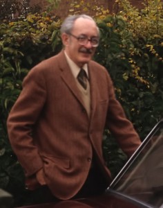
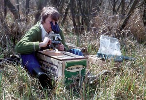
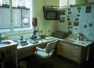
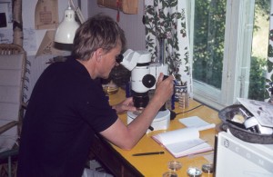
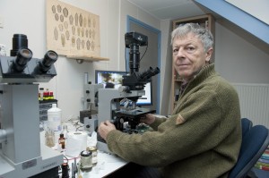
Ferry Siemensma at his Microscope

Ferry Siemensma at the microscope

Ferry Siemensma is an independent researcher in the Netherlands, and a leading expert on several groups of amoeboid and heliozoan organisms. For the past two years he’s been building a website devoted to his favourite creatures: Microworld: world of Amoeboid Organisms. This was an ambitious project from the beginning, but over time it has gradually turned into something that hasn’t been seen since the comprehensive texts of Cash, Wailes and Hopkinson, or Eugène Penard: a wide-ranging illustrated guide, featuring good formal descriptions, keys for identification, and up-to-the minute taxonomy. Ferry kindly agreed to answer my questions, in the first of what I hope will be a regular series of “Meet the Protistologist” interviews.

Your enthusiasm for your research reminds me of Joseph Leidy’s recorded remark: “How can life be tiresome so long as there is still a new rhizopod undescribed?”   He was not raised in natural science (his father was in the hat business), but even in childhood he was driven “to see how plants and animals are made.” What was your upbringing like?

I grew up in a rural community in the northern part of the Netherlands. My father was a carpenter and I was the oldest of nine children. We always played on our uncle’s farm around the corner, and there I got my first interest in nature. I became a voracious reader of popular books about science and biology and in one of them I read the word “amoeba”. For me it was such a magic word, I don’t know why. From then on I was determined to find such a creature once. I was 14 years old when I bought a cheap Japanese microscope for about $10, which was pretty expensive in 1962. I could recognize some moving protists with it, but no amoebae. Many years later, at the age of 22, I became a teacher in a primary school, where I worked with 11-12 year old children. I also took a study to become a biology teacher and during that study we were offered to buy a Lomo microscope. This Russian instrument was really great, and it had a wonderful 70X water-immersion. From the day I got that scope, my life changed completely!

So, you were a schoolteacher, like Alfred Kahl! What changed in your life after you acquired the Lomo?  

From then on, for more than 12 years, I looked through the microscope nearly every evening. My three daughters were very young at that time and went to bed very early, which was a great advantage ;-). Very soon I got in contact with Rob Wellner, who was looking for a microscope friend. We worked together for two days a week for many years. He taught me a lot, but his main interest was algae and I shifted to amoebae and heliozoa. In that time I got in contact with Dr. De Groot, a well known Dutch amoeba specialist. We smoked a lot of cigars together and had nice talks about our shared interest.

1980-Fred-Page-Cambridge

Frederick C. Page at Cambridge, 1980 (Source: Ferry Siemensma)

When he died in 1981, he left me his scientific literature, with the books of Leidy, Penard and Cash, which was a great luck for me, because in that time it was very difficult to get any papers. There was no internet, which is very hard to imagine now. In the meantime I also got in contact with Fred Page in Cambridge. He is such a pleasant man and we wrote each other a lot of letters. I went twice to Cambridge to see him.

He gave me the address of a Danish microscopist, Niels Willumsen. I phoned him, he invited me and that was the beginning of a very long friendship. One day I got a letter from Rudi Roijackers, who worked at the University of Wageningen. He offered me the use of an electron microscope and that was an excellent chance to get new information about heliozoa. I hunted for heliozoa, isolated them, made dried preparations and once a week we looked at the results in the SEM. We discovered new species and published some papers. I also published two booklets in Dutch about heliozoa and naked amoebae. Than I was invited to write the heliozoan part of the German Protozoenfauna. Fred Page wrote the other part on naked amoebae. The book was published in 1991.
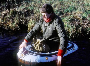
1981-Ferry---Sampling-in-Kortenhoef

Ferry on a sampling excursion, 1981. (Photo by Niels Willumsen, who later capsized in the same boat)

Looking at your publications during this period, you were very active, especially with heliozoans. It must have been a thrill to be among the first to see EM images of the very intricate scales in certain groups! Were there certain organisms that you found particularly fascinating?

1982-Field-study-with-my-Lomo

Ferry in the field with his Lomo, 1982. (Source: Ferry Siemensma)

I was intrigued by the scales of Rabdiophrys species, though they aren’t true heliozoans. Those scales had been studied ten years before by Thomsen and I found them very beautiful subjects to make drawings of. One problem was to interpret the information on the SEM photomicrographs and to form a 3-dimensional image of those scales, which was really a challenge, and very fascinating, which was also true for the base of many kind of spiculae of the true heliozoans.

If you don’t mind my asking, was it a struggle to find funding and free time for this research?  Did you continue to teach?

No, there was no funding, because the work at the university of Wageningen was part of the research of Rudi Roijackers. His interest was Mallamonas and he wanted to find out which scales belongs to heliozoans and which to Mallamonas species. He helped me with the SEM, I helped him with the identification.

Concerning my free time, I did all my research in the evening, the weekends and in my ample holidays. The latter was the big advantage of being a teacher! Schools were closed on Wednesday afternoon, so once a month on Wednesday I jumped into my car and drove to the SEM, one hour driving from my house! Teachers didn’t have a great salary, and they still don’t, so in that time I cleaned every cover glass in order to reuse it 😉

I still clean coverslips, unless they have immersion oil on them!:) In the decade between Nackte Rhizopoda und Heliozoea and the ISOP Illustrated Guide, you seem to have published less frequently. Were you pursuing other interests, or simply working quietly on your own?

1984-Looking-at-Penards-slides-in-the-British-Museum

At the British Museum in 1984, to look at Eugène Penard’s slides. (Source: Ferry SIemensma)

There came a moment that I didn’t find any more new heliozoans. Preparing them for the SEM was a lot of work. First I had to find one, then to decide if it could be a new species, then came the process of removing the cover glass, which was the most complicated part, isolating the specimen, drying it, marking it and finally cutting the slide for the SEM.

After some months without any positive hit, my interest faded. It was at that time, as my girls grew older and needed my attention, that I got very interested in computers and I had a chance to start a science magazine for children – all in my free time. For many years a layer of dust grew slowly on my microscope…

Did you make room for amoebae in your magazine?  It always seems to me that popular science publications have too many telescopes and not enough microscopes!

No, it was intended for 10-14 year old children, and they don’t usually own a microscope.

When did you decide to blow the dust off your microscope, and what brought you back?

Some years ago I got a letter from a woman working at a waterlaboratorium here in the Netherlands. They control the drinkwater quality and had problems with amoebae transporting Legionella bacteria through their filters. They needed my help for identification and had found my name with Google. And there I went to the lab and worked a whole day with a microscope…after so many years. I enjoyed it so much, it was a wake-up call. From that day on, I decided to start again. My first look through my Russian microscope taught me that it was unusable anymore: completely worn out. I bought a secondhand Zeiss standard and later an Orthoplan with DIC and phase contrast and other stuff on eBay. And so I came back home again!

In your website, your experience as an educator and your expertise with amoeboids have come together. How did this project begin?

I started this project about two years ago with the intention to make a survey of all the Dutch amoebae, and as replacement for the book of Hoogenraad & De Groot (1940). Here I could combine my interest in computers, nature, photography and drawing. But soon there came questions from people in the forums who asked for identifications of their findings and I started to publish their photographs also on my site. And then I realized it would be nice to cover the whole world. But it’s a huge project, in fact too much for one person. Every day there are new discoveries to document, new photomicrographs to edit and to archive and new email to answer. But I’m building my site stone by stone… uh.. shell by shell, without hurry and without any pressure. I love it!

While developing your website, you are continuing to do original research.  Can you talk about that work?

1982-Niels-Willumsen-in-his-summerhouse-in-Zweden

Niels Willumsen at his country home in Sweden, in 1982. (Source: Ferry Siemensma)

I’m very interested in the variation within populations. Probably you know how difficult it is to distinguish between certain Centropyxis species, and the same is true for Difflugia, Euglypha, Cyphoderia and many other taxa. I’m measuring populations from different localities and will see what comes out. At the moment I have an Austrian sample with a population of Pseudonebela africana, which is new for Europe. It has been described from Africa and Brazil as having dimensions between 78 and 100 µm, but I find shells which are 85-185 µm long. That’s really interesting, and shows how little we know of populations in the wild. As these specimens are living, I also have the opportunity to observe their feeding behavior which is the same of that of Difflugia rubescens, and these specimens also have the same red color. They must be related. I’m fascinated by the behavior of amoeboids. I’m using a nice and simple, but very successful method for observations, the so-called “wet chambers” where I keep my wet mounts. It’s always a surprise, what comes up in such mounts after several days. Not seldom some species multiply rapidly and attach to the cover glass. which is excellent for observations. Mmm… I can go on for many hours…

It would be a pleasure to spend those hours with you, and I hope I will have the chance, one day.  Thank you so much for taking the time to do this.
Dec 082013
 

I’ve added a number to the title of this post, because I expect to make “Protist Homes” a regular feature. I had intended it to be an idea-free zone, devoted to uncomplicated wonderment (kind of like the tours of celebrity homes and stately residences on HGTV).  But I know some ideas and research will creep in, because it’s hard to feel wonderment without actually starting to wonder.

Difflugia acuminata

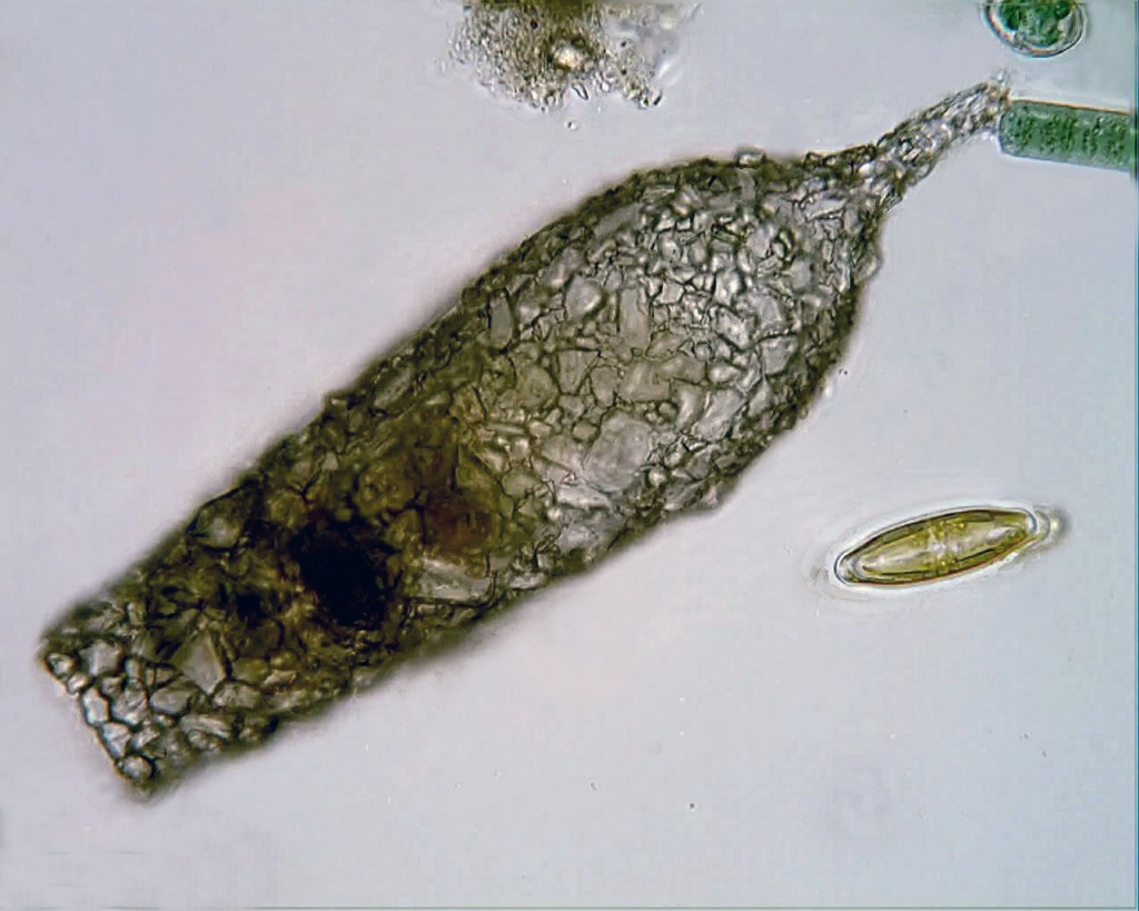
Difflugia acuminata, from a pond in Wakefield, Quebec


Consider the little shell in the image to the left.  Barely two tenths of a millimetre long, it was constructed by an amoeboid, using found materials: tiny grains of sand, glued together with some sort of proteinaceous cement.

How does a single-celled amoeba build such a perfect bottle, from such randomly-shaped material?  And why did my grade-school macaroni art projects turn out so much worse?


The organism that once lived in that test is dead (testate amoebae cannot leave their shells without dying). In life,  it would have looked and behaved like the one in this video, by my fellow protist-watcher, Francisco Pujante:

All members of the genus Difflugia build their shells–or tests, as they’re properly calledout of stuff they find lying around, such as grains of quartz or discarded diatom shells, binding them into a matrix of organic secretions. It turns out that they are quite selective about the particles they use for this work, and researchers have gone to a lot of trouble to figure out what criteria amoebae use when picking their building materials.

Difflugia 3D rendering 2

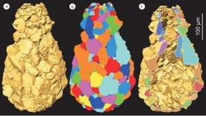
3D rendering of Difflugia oblonga, created by Châtelet et al. Click image to show source.

Recently, a group in France went so far as to create complete three-dimensional reconstructions of two specimens of Difflugia oblonga, using a process called x-ray microtomography. They calculated the volume, shape and orientation of each grain in their specimen tests, and gathered information about the density and composition of the individual particles. Yes, you read that correctly: they analyzed and modelled every micro-sandgrain in amoeba shells that are themselves smaller than the grains of sand you might find on an ordinary beach.

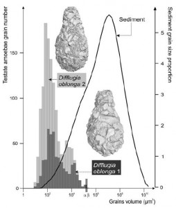
Using all this data, they compared the composition of the tests to the natural sediments in the ponds where they were collected. From their analysis, it appears that these amoebae really are selecting the smaller grains, and also showing a taste for certain minerals (quartz is preferred, while the more abundant calcite is shunned). What’s more, the amoebae seem to be selecting materials with a consistent density, which the authors speculate is to ensure the test will be perfectly balanced!

Difflugia grain sizes


The paper (du Châtelet et al, 2013), includes a handy graph, comparing the sizes of each of grain of sand in two Difflugia tests to a distribution plot of the size of sand particles in the surrounding pond sediment.


Building with quartz is fine, if you are lucky enough to live in the mud bottom of a pond where chunks of silicate minerals are available.  But what if your habitat is a floating clump of algae in a mineral-poor spagnum pool?  There, the best source of silicate particles might be the siliceous shells (frustules) of your fellow protists the diatoms, which have the ability to build their little glass canoes from dissolved silica (silicic acid) found in the water around them. The testate amoeba Difflugia baccillariarum–typically found in sphagnum pools, where sand is in short supply–has figured out how to glue diatoms around itself as a means of protection.  I find these guys, sometimes, in samples from the Mer Bleue bog,  and always stop to look at them.  They are truly amazing:

It is hard not to admire this crafty amoeboid, building its home from the bodies of other organisms.  Sitting here in my post-and-beam house, assembled from the trunks and branches of felled trees, I feel a certain kinship.


Dec 062013
 

The Last Eukaryote Common Ancestor–affectionately known to cellular evolution geeks as “LECA“– was almost certainly a predator.  Later in the history of the lineage, certain eukaryotes would form permanent symbiotic alliances with photosynthetic bacteria, and once that occurred, some lucky cells could just bask in the sunshine, drawing energy from light with the help of their chloroplasts. Until that happy day, however, all nucleated cells survived by using a bizarre trick that LECA must have known: the ability to ingest other cells by phagocytosis (literally, “cell-devouring”).

Another thing about LECA and its descendents is that, thanks largely to the mitochondria which give them a fairly generous energy budget, eukaryotes of all kinds have a real genius for evolving new and outlandish bodily structures. Unlike the prokaryotes, bacteria and archaea, which tend to have rather unimaginative body shapes, energy-rich eukaryotic cells have developed a huge (and hugely entertaining) diversity of organs, appendages and specialized equipment.

So, in the eukaryotes we have a population of ravenous cell-eating cells that also have a talent for morphological innovation. Clearly, this has the makings of an arms race, as prey and predator (and most eukaryotes are both!) hurry to develop new instruments for attack and self-defense. This race  is one of the likely drivers of their exceptional somatic diversity. There are other evolutionary drivers of eukaryote complexity–including some not directly tied to adaptive pressures–but the need to avoid being devoured  is certainly an important one.

There are many ways to avoid being eaten. One of the simplest strategies is to be too big to swallow. However, there is a cost to that.  Big bodies are expensive: they require a lot of energy to keep going, which means the organism has to gather more food.  But what if you can be big without having to maintain a large mass of living tissue?

One way to accomplish that is to congregate with others of your kind in close-knit (even clonal) colonies, as many flagellates do.  Take, for example, the planktonic heterokont Synura, which usually lives in big, rolling spherical clusters made up of many cells. If you want to eat a single Synura, you will need to be big enough to eat the whole sphere.

Obviously, there are plenty of creatures big enough to consume a whole colony (I’ve seen Stentor pyriformis gobbling Synura like popcorn balls); but the strategy of colonial aggregation does take some of the smaller predators out of the game.

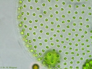
There are a few downsides to colonial living (decreased freedom of movement, for one); but the tradeoffs can be acceptable, especially for those protists whose way of life doesn’t require much personal mobility. Not surprisingly, photosynthetic organisms, which don’t have to chase their dinner around, often live in colonies.  In the microworld, familiar examples are the filamentous green algae, or the great spherical death stars we call Volvox (which are not only colonial, but actually have some cellular specialization, like plants and animals).

Volvox by R. Wagner

Volvox aureus. Image by R. Wagner. Click on the picture to see more Volvox on Dr. Wagner’s site.

Another good survival strategy is to borrow “bigness” from your surroundings.  Mix yourself in with a pile of debris, or crawl into a hole in the mud, and your enemies will be unable to get their mouths around you.  In short: take shelter.  It is the same plan, here, whether you are a protist, a beetle, or a juicy human surrounded by bearsharks: position yourself inside something bigger, pricklier and tougher than your own delicious body, and you might survive.

One major disadvantage of opportunistic shelter-seeking, as practised by cautious organisms of all sizes, is that your fortress isn’t always located close to your preferred food source; and if you leave it to go foraging, you risk becoming a food source yourself. One good solution is to build (or secrete) your own shelter in an optimal location. This approach, often combined with the strategy of colonial living, has given rise to an amazing array of ingenious shells, houses, tube dwellings and domestic mucous piles which protists have created for their own protection. In my next couple of posts, I will look at some examples of protistan architectural achievements.

Nov 292013
 

As I was saying, we vertebrates tend to have a high opinion of the big clump of neurons that sits at one end of our spinal cord.  And so we should: in creatures like us, that’s where the magic happens.  The bigger the clump, the more cognitively versatile the organism; remove it, and the whole organism comes to a stop (though it may run around for a while, first).

But, as we know, even unicellular beings are capable of sensation and coordinated movement, and can show complex, even purposeful, behaviours.  So, it was probably inevitable that some well-meaning scientist would try to equip them with a central nervous system of their own.

Stylonychia crawling Actinophrys

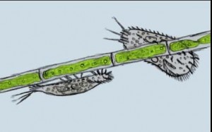
Stylonychia mytilus crawling on a filament of algae. Image by Actinophrys, on Flickr. Click for Link.

In 1880, a German researcher named Th. W. Engelmann undertook a close inspection of the tissues of the ciliate Stylonychia mytilus.  Like other members of the ciliate group loosely known as “hypotrichs,” Stylonychia has, on its “belly,” a crop of thick, mobile cirri: bundles of fused cilia that it uses like little legs, as it scrambles around on solid objects in its sunken world.  These pseudo-legs work very well, and a crawling hypotrich can look a lot like a beetle or cockroach.

Unlike the limbs of insects, the cirri of hypotrichs lack conspicuous attachments, such as muscles, tendons and nerves. However, looking very closely at stained samples, Engelmann was able to detect, at the bases of Stylonychia’s cirri, groups of fibres which he called “Wimperwurzeln,” or “ciliary rootlets.” These fibres, he speculated, might just function as nerves do in “higher” organisms, serving as the conductive tracks along which the stimuli that move the cilia might move!

It was an innovative idea; however, it did not spark much excitement. Despite his impressively clear record of these small structures, the suggestion that he had discovered animal-like organs in unicellular creatures might have come at the wrong time. By 1880, the view of protozoa as miniature animals, bearing a complete set of reproductive and alimentary organs, had been discredited a long time before. In the late 19th century, protozoa were conventionally seen as primitive organisms, little more than bags of the all-purpose protoplasm that constituted, in Thomas Huxley’s memorable phrase, the “physical basis of life.”

But times change, and the idea that free-living cells might be composed of differentiated tissues would came back into style (in fact, in the early 20th century some protozoologists completely rejected the cellular nature of their organisms).

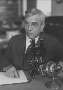
Charles Kofoid picture

Charles Atwood Kofoid

In 1914, Robert G. Sharp–working in California under the guidance of the eminent protozoologist Charles Atwood Kofoid–published a paper in which he boldly claimed to have found a ciliate that not only had nerve-fibres, but a full-blown centrally located “neuromotor apparatus.” He discovered this structure in an organism called Diplodinium ecaudatum (now known as Epidinium ecaudatum), a symbiont in the stomachs of cattle. What Sharp found had never been recorded anywhere: a small mass of fibres close to the cell’s motile organelles, and serving as “the common center of motor influences.” He had discovered the organ that controlled and coordinated the moving parts of the ciliate, in effect, the cytoplasmic “brain” of the cell. He called this the “motorium.”

Sharp’s paper kicked up a little whirlwind of scientific activity, particularly among his colleagues at Kofoid’s laboratory. The following year, Kofoid himself described a “neuromotor apparatus” in the intestinal parasite, Giardia. Shortly after, Harry B. Yocom, another worker in Kofoid’s group, published the discovery of a full-blown neuromotorium in the hypotrich Euplotes patella (a “walking ciliate” like Stylonychia).

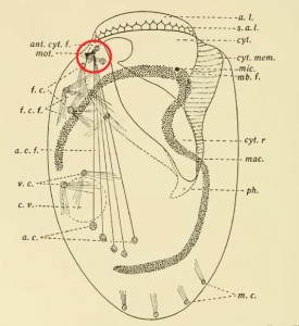
But why ascribe a neural function to the structures he saw in Euplotes? Yocom goes into some detail about that. In the first place, these fibres turn a vivid red in the fuchsin from Mallory’s stain, just as nerve cells do in metazoa; in the second place, he explains, these fibres are found in association with the moving parts of the ciliate: the oral membranelles and the locomotory cirri.

Yocom Euplotes neuromotor apparatus circled in red

Yocom’s illustration of the neuromotor apparatus in Euplotes patella, with the “motorium” circled in red.

Finally, the behaviour of Euplotes patella appeared to support this interpretation. It is, perhaps, a classic example of hypothesis leading observation by the nose. To Yocom, it seemed evident that E. patella was using its “anterior lip” (the bulge at the top of the cell, which showed a red-staining lattice of fibres) as a sensory organ. What’s more, the transverse cirri, which were linked to the “motorium,” moved in a coordinated way, while the unattached cirri moved randomly.  As Yocom puts it: “[T]he whirling irregular movements of the frontal ventral and marginal cirri are in no way coordinated with the regular rhythmical movements of the membranelles or with the backward kick of the cirri.”

Or, so it looked to him.

Up to that point, the function of the “neuromotorium” as a “coordinating center” for the cell’s movements was simply a hypothesis, unsupported by experiment. But confirmation was not long in coming. In 1919, C. V. Taylor–yet another worker from the Berkeley circle–conducted an experiment on Euplotes patella, the ciliate Yocom had studied in such detail the year before. Using quartz microscalpels, he severed the fibres running between the motorium and the anal cirri (the prominent group of five heavy cirri in the posterior half of the cell, now usually known as “transverse cirri”). The experiment resoundingly confirmed the hypothesis; detached from the motorium, the cirri did not seem to work properly at all: “Severing the fibers to the anal cirri affects both creeping and swimming…Destroying the motorium or cutting its attached fibers interrupts coordination in the movements of the adoral membranelles and anal cirri.”  (Taylor, 1919)

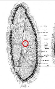
Paramecium nerve center with red circle

Paramecium from Rees (1920), showing a tracery of fine “neurofibrils” (skeletal microtubules, presumably) converging on the “nerve center” of the cell, circled here in red.

From then on, the neuromotor apparatus turned up in one ciliate after another. It was found in Paramecium (1920), Balantidium (1922), Tintinnopsis (1926), Dileptus (1927), Chlamydodon (1928), Uroleptus (1930), and Oxytricha (1935). Along the way, doubt steadily faded, and by 1927 it was possible to affirm plainly that “a complicated neuromotor system has been conclusively demonstrated to exist in certain ciliated infusoria.” (Visscher, 1927) It had become, in North America, at least, a solid fact.

But even solid facts can turn fluid again, and then evaporate like the canals of Mars.  It can happen slowly, or all at once.  Sometimes a hypothesis dies a quick death, when an experiment disproves it; sometimes it is swept off in the riptide of a better theory; and sometimes it just get nibbled away, while the world slowly changes around it, until one day nobody can remember why it had ever seemed true.

The neuromotor concept had a long run, outlasting Kofoid himself by a decade and a half.  Even the advent of electron microscopy did not kill it off, right away.  As late as 1957, EM seemed to reveal in Euplotes “a mass of intertwining rootlet filaments” corresponding to the neuromotorium, as well as a whole system of intracellular fibres which were mainly dedicated, as the authors argued, to “the coordination of the ciliary beat.” (Roth, 1957) But when R. Gliddon again placed Euplotes under the electron microscope, in 1966, he could not find anything you could call a motorium.  It had disappeared.

In that same year, two researchers in Japan finally got around to replicating Taylor’s classic experiment on E. patella–the experiment that, arguably, had kicked off the neuromotorium gold rush. (Okajima and Kinosita, 1966) But now, the results were very different.  The investigators severed the “neurofibrils,” just as Taylor had done, and captured the ciliary movements on film; however, this time the cirri just kept on moving in their usual coordinated way, as if nothing had happened. It is not known why Taylor’s Euplotes had behaved differently.  Possibly, he mangled the poor things while dissecting their “neurofibrils” and made his celebrated observations on organisms in their death throes.

In 1970, Dorothy Pitelka was ready to call the game, announcing the general “abandonment of the old concept of a fibrillar neuromotor system in ciliates.” A casual search in Google Books turns up one last, brief reference to the “neural fibrils” of Paramecium, in a high school biology textbook published in 1983.  After that, nothing.

So what were the “neurofibrils,” in the end? They were not a product of poor equipment or fanciful observation, but rather the opposite. Remarkably, it seems that microscopists of the day were already detecting parts of the cytoskeleton–the scaffolding of microtubules and filaments that give the cell its shape–as well as the pellicular “latticework,” the so-called ciliate “silverline system” which would later be fully exposed in silver nitrate and protargol preparations. Interpretation lagged behind observation for a generation or two. Seeing had outstripped understanding; then new information came, and the old certainties just sank back into the pondwater.

Click Here for a List of References

Nov 232013
 

When René Descartes searched through the human brain to find the Seat of the Soul, he settled on the pineal gland, a small endocrine organ found in nearly all vertebrates. It seemed like a good candidate, because it is a singular organ, not duplicated on each half of the brain but perched right on the midline, where it could, as Descartes imagined, receive sensory impressions through little pipes, and flex the muscles of the body by exhaling “animal spirits” along a system of hoses leading to the arms and legs.

Descartes brain 4b

Illustration from Traité de l’Homme (1644), showing the teardrop-shaped pineal gland perched in the skull like a little Mecha-pilot.

Descartes’ soul sits at the control centre of the meat-puppet, gathering news of the world through its eyes, ears and nose and exerting its will upon the world through the pneumatic machinery of the body.

It is easy to recognize in his conception a structure analogous to  what we know as the nervous system. We no longer think in terms of “animal spirits,” but rather of “neural signals,” and we no longer place the mind within any particular gland in the brain. But for most of us, who have only a rough understanding of how it all works, this “signal/transmitter/receiver” scheme is only another metaphor, just a little more sophisticated than the one Descartes used. By and large, we are still operating with the notion of an insubstantial mind somehow localized within a “central” nervous system, conducting its business with the world through an extensive network of  bodily filaments.

Indeed, many of us find it hard to even conceive of an intelligence which is not concentrated  at the top of a hierarchy, running from the brain (where the cogitating self resides) to the tips of the peripheral nerves, where the world lies waiting to be felt, heard and seen.

So, the distributed intelligence of the octopus nervous system–half of its neurons are in its arms!--strikes us as something bizarre and wondrous, requiring further exploration and explanation. Challenging the naive expectation that cognitive processing should be “central” and the organs of sensation “peripheral,” the octopus researchers have devised ingenious experiments to show that the arms of the mollusc continue to perform goal-oriented behaviours, like prey-seeking, exploration and danger-avoidance even when they are severed from the so-called brain.

And if it seems marvelous that a detached octopus arm can still sense and react  to perceived danger, how much weirder is it that a single cell can do all these things, without the benefit of any nervous system at all? But this is perfectly commonplace in the protist world, where one-celled hunters swim around, stalking one-celled victims, who recoil when they sense trouble, take shelter in their shells, or hide from their enemies in clumps of debris.

Just imagine if a single cell of your body could cut loose and go wandering about under its own power like that, hunting and consuming prey! Oh, right…they do. In fact, it is perfectly ordinary for eukaryote cells, whether they are inside larger organisms or roaming at large in the “world,” to exhibit complex behaviours that may be remarkably hard to distinguish from the kinds of behaviours we observe in large, multicellular animals.

Take a ciliate like Stichotricha aculeata. It usually inhabits a shabby, homemade tubular dwelling (a lorica), made up of mucus mingled with debris. It lives by brushing smaller organisms — bacteria and some of the daintier protists — into a mouthlike aperture, where they are packed up in little food-bubbles (vacuoles) and digested. Like a cat or a crayfish, it is both large enough to be a predator, and small enough to be prey. Consequently, it is a cautious creature, relying on its lorica for protection. When it senses trouble, it withdraws into its tube, and when the coast is clear it will put out its long feeding apparatus. Like other ciliates, it can sense movement (probably with the help of sensory cilia, which are common in eukaryotic cells), and is easily startled. If you should tap on the microscope stage with your finger, while you are watching it, it will detect the vibrations and pull back.  After hiding for a while in its jelly-tube, it will extend its proboscis, tentatively. If it encounters no more trouble it will begin to feed again.

However, if the disruption is sufficiently violent or prolonged, it may abandon its lorica altogether, as the closely-related Chaetospira does in a video I recorded last year. The Stichotricha in the embedded footage which follows has already fled its original “home,” and taken shelter in a little enclosed space, between the coral-like tubular dwelling of a colonial flagellate called Rhipidodendron huxleyi, and a mucoid colony of some other organism (Spongomonas?). In this makeshift lorica-substitute, it is behaving exactly as it normally does when at home, sliding forward and back, feeding, withdrawing, advancing, always hungry, and always vigilant:

It is easy to see why early microscopists referred to these creatures as “animalcules: “very small animals”. Without a nervous system, ciliates like Stichotricha and Chaetospira exhibit behaviours strikingly reminiscent of a fish lurking in a crevice, or a case-dwelling insect, like the larval caddisfly. If they often remind us of “true” animals, it is well to remind ourselves that, after all, they have a way of life that differs very little that of other cavity-dwelling aquatic creatures, like the ones we might find on a coral reef, or like this larval midge, in the gravel of a freshwater stream:


Nov 222013
 

Stewart Brand usually gets credit for the quote, but apparently it was Craig Venter who said it: “If you don’t like bacteria, you’re on the wrong planet.  This is the planet of the bacteria.”

Actually, the second part of that, the play on Planet of the Apes, was probably lifted from the title of an article by Stephen Jay Gould, included in his book Full House.  When I was asked to review the book, eighteen years ago, one passage blew a hole in my world view: “We live now in the ‘Age of Bacteria.’ Our planet has always been in the ‘Age of Bacteria,’ ever since the first fossils—bacteria, of course—were entombed in rocks more than 3 billion years ago.”

It’s funny that it was Stephen Jay Gould who steered me away from my zoocentricity, because the preeminent explainer of evolutionary biology rarely had much to say about microbes. Pulling one of his books off the shelf and scanning for organism names in the index, I see: ammonites, aphid, angler fish, Archaopteryx, asses, Australopithecus–ah, there’s “bacteria,” with two brief entries–and then, bees, birds, Blatella germanica (a cockroach), blue-footed boobies, boobies again, then brown boobies, brown hyenas, coelecanths….and so it continues until we get to Eschirichia coli, and much later, two entries for “prokaryotes.”  That’s it for microbiology in that book (Hen’s Teeth and Horse’s Toes).

Protists don’t rate a single mention. And it is much the same story in Gould’s other works.  Even his desk-bending opus ultimum, The Structure of Evolutionary Theory, mentions “protistans” just a handful of times, mostly in connection with the observation that gradualism will govern the rate of evolutionary change in “asexual” organisms.  (Most protists are as sexy as can be, but let’s leave that aside, for now).

I didn’t bring this up to bash Gould.  He wrote mainly for a general readership, and microbiology wasn’t his thing anyway. But perhaps it reveals something that, as late as 2002,  it was possible to write a 1,400 page treatise on evolutionary theory in which the only source cited on “protistans” is D’Arcy Thompson’s On Growth and Form, published in 1917. And I’m not sure that Gould’s zoocentrism is all that unusual in his field.  Certainly, Lynn Margulis (enthusiastically wrong about some things, but the best friend a microbe ever had) often complained loudly about the dominance of “zoologists who today call themselves ‘evolutionary biologists’.” (Acquiring Genomes, 26) Last year, there was an international congress on evolutionary biology in the big city down the road from my village. Scanning the program, I noticed that most of the talks were centred around macroorganisms of one kind or another (tigers, termites, toadstools, and tumtum trees). A friend who teaches evolutionary biology at CUNY was in town for the event. “Don’t worry,” he teased me, “there’s bound to be one or two people giving talks about the weird stuff!”

As my friend knows quite well, here on the “Planet of the Bacteria,” the “weird stuff” is really creatures like us: great, shambling, genetically-coordinated quasi-colonies, lurching around with more than 37 trillion specialized cells inside them (and hosting perhaps ten times that number of hitchhiker microbes).   The protists seem almost normal, by comparison, though even they are evolutionary oddballs.  All of us eukaryotes are weird, but, some of us are weirder than others.  While plants, fungi and animals make up only a few remote twigs at one end of the eukaryote lineage, the long sideways-projecting stalk on which they sit– comprising what is sometimes called (though I can hardly say it without snickering) “Empire Eukaryota”– is made up mainly of (royal fanfare, please) “Kingdom Protista.”

Needless to say, the natural history of eukaryota was mostly written by protists, and by all rights the study of them ought to be central to evolutionary theory.   Bear in mind that if the original ancestral eukaryote were to emerge from some magic time capsule and land in the petri dish of a modern biologist, it would almost certainly be classified as a protist.   And, if you look closely enough at some of our 37 trillion cells, our kinship with them is hard to miss.  Our lungs and fallopian tubes have genuine cilia that beat just like those of the ciliated protozoa.  Our sperm have flagella (really just another kind of cilium), no different, structurally, from those that power any flagellated protist.  Our bodily fluids are patrolled by ravenous amoeboids that roam around, engulfing bacteria and other other invaders.    Out here in the “Empire” of nucleated cells, protists are us.