Dec 092014
 

Eckhard Voelcker (right) with Steffen Clauß

Its been less than half a year since Eckhard Voelcker and Steffen Clauß launched their enchanting online gallery of amoeboid organisms, www.Penard.de. Already, it has emerged as one of the best places to find micrographs of amoebae and heliozoans, and the collection is continuing to grow and improve. The site features some superb light microscopy, but also sumptuously clear and detailed scanning electron micrographs prepared in Eckhards own basement laboratory.

Eckhard is an unusual guy.  A self-taught programmer, he spent several decades in software development (at the head of Völcker Informatik AG) before turning, in midlife, to the exploration of amoeboid protists. He has pursued the new discipline in a remarkably organized and self-assured way, and, in collaboration with Steffen Clauß, has begun to make real contributions to the field. As an amateur scientist working at a high level, Eckhard seemed like a perfect subject for Meet the Protistologist. I contacted him a few weeks ago, and he agreed to answer some questions.

“The child is father of the man,” as the poet Wordsworth said. I would like to know something about your earlier life. Where did you grow up? Were you exposed to the natural sciences as a child?

I grew up in northern Germany. Natural sciences were always my favorite subjects and I wanted to study chemistry or biology. Somehow I ended up becoming a computer scientist and moved to Berlin. Here I started my own software firm a couple of years later. Biology became a hobby of mine and I enjoyed watching microbes with the microscope to relax after work. It was in the year 2010, after a large corporation bought my firm, that I started to think about my future. So I started to plan for an early retirement and went as often as possible to the university, where a friend of mine, Klaus Hausmann, was a professor teaching and researching protistology. After Klaus introduced me to the electron microscope, I was hooked. I planned and built a protistology laboratory with an electron microscope in my basement and decided to focus on this and not software anymore.

So, you had no formal training in protistology or microscopy, before you began to visit Klaus Hausmann’s lab at the Freie Universität Berlin?  Did you do any work at his facility, or were you simply observing?

I had been a member of the Berlin Microscopic Society for some years and my microscopic interest was mainly protists. So I did spend many hours observing, reading papers and studying books. When I would find an amoeba unknown to me, I would try to identify at least the genus and then read the available literature about it. That was why I started with the EM in the first place. I found Cochliopodium amoebae and was interested in the scale structure that is invisible with the light microscope. Somehow I convinced Klaus Hausmann that this would be interesting and he promised to help me learning the EM preparation and operation. There was this magical day a couple of weeks later that I remember so well. I had brought a culture of Cochliopodium vestitum to the university and we put some cover slips in petri dishes and filled these with my culture. A couple of days later it was my birthday and I had decided to take the day off and prepare for the EM. In the afternoon, when the preparation was done, I went to Klaus’s office proudly holding 6 sputter-coated coverslips on EM stubs and together we went down to the SEM. It was a magic moment when I first saw those delicate scales – and it was my first very own preparation (Klaus said I was very lucky to be successful right away – oh boy, was he right). When I had dinner with my wife that evening, I told her, this is what I want to do when I am done with my job. It took me some years though to finalize my work and to build my lab, but now it is all done. Klaus Hausmann has retired two years ago and the lab at the university is not a hotspot for protistology anymore as his successor is focusing on evolution.

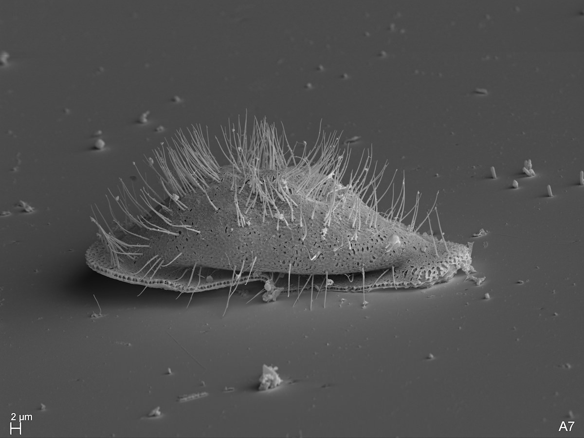
Cochliopodium vestitum (source: Eckhard Voelcker).

You’ve mentioned to me that you learn best on your own, outside an academic context. Could you elaborate on that?

It has nothing to do with the academic context. When I listen to a lecture on video, I will pause that lecture a dozen times or so and check something, often to dig deeper into a certain detail. After my curiosity is satisfied, I will continue the lecture. I cannot do this when somebody stands in front of a blackboard “live”.

How did you begin planning your home lab?  You must have been acquiring new knowledge and skills at a prodigious rate.

Planning my lab was fairly easy. I knew what I needed for preparation and there was only one room available anyway. So basically it was just the question, how do you fit a fume hood, refrigerator and deep freezer for chemicals, a SEM, sputter coater and critical point dryer into one room. Originally I planned to leave the light microscopy at my old lab in my study, but this proved to be not practical. So I moved everything down into the lab, although it is a bit cramped now. But as my wife said, this removed the danger of me buying yet another microscope.

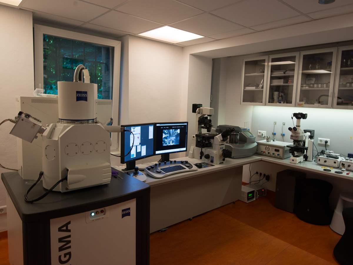
Eckhard’s home laboratory, with Zeiss Sigma Scanning Electron Microscope at the left. (Source: Eckhard Voelcker)

Did you have collaborators in the beginning?  Were you already working with Steffen Clauß?

 I got in contact with Ferry Siemensma pretty early. He helped me to identify some amoeba and we have spent many hours on the phone. It was Ferry who brought Steffen and me together. Steffen had found an undescribed testate amoeba and wanted to know how to prepare it for the EM. Eventually we met and got to know each other. We were in the same position, hobbyists working alone with an exotic (to say the least) passion for amoebae. In the spring of this year we came up with the idea to create a website about amoebae combining light- and electron-microscopic images.

So, even at the start you had a particular interest in amoebae. What attracts you to them?

When I had my first small microscope it did not take long to stumble across Arcella discoides. It looked so strange, so perfectly round, simply incredible, like a flying saucer. I realized that there was more to amoebae besides being a blob of slime. The more I looked into amoebae and heliozoans the more fascinated I became.

Have you singled out certain taxa or biological phenomena for closer study? 

I have a special interest in cells with scales. Some of the “naked” amoebae are really not naked, but covered with minute scales of a specific structure. Korotnevella and Cochliopodium are two nice examples. Also some heliozoans, especially Pterocystis fascinate me. When we sample in unspoiled environments, we will find species that have been more or less forgotten since over 100 years and that have never been imaged with an electron microscope. We also find many undescribed species. When I load a preparation into the electron microscope and I see an undescribed species, it really is some kind of golden moment. I am the first human who is seeing this species. But the challenge is to isolate and culture so molecular data can be obtained. Without molecular data you cannot describe new species these days. And publishing what we find is an important goal. I hope that next year we will be able to do this.

Korotnevella sp. looks like a “naked” amoeba in transmitted light, but SEM reveals its cloak of intricate scales. Source: Eckhard Voelcker.

Many of your micrographs are as aesthetically pleasing as they are informative.  Do you have any thoughts about the role of art in science, or science in art?

This is a very interesting question. Most boys of my generation, as some generations before me, received at some point in their youth a microscope as a present. Looking into pond water to see the wonders of paramecium, euglena etc. was very common. Today internet gaming, social media etc. seems to be more popular and „young scientists“, who explore their surrounding nature with the microscope or a looking glass have become very rare. If we want to make people understand, that environmental protection is not only about rare plants and animals, but about unspoiled environments, swamps etc, we need to show them there is something wonderful living in these waters. If we want to make people excited about science, we need to make it visually attractive. I fully understand why most scientists don’t bother about aesthetics in their images. They have little time, are under pressure to produce papers etc. But this will create little public enthusiasm about their work. And this leads to less funding. For Steffen and me, if we can’t show an amoeba in a nice image, we won’t show it at all.

In many natural sciences, even in mathematics, very often the most fundamental equations are very aesthetic.  Look at the mandelbrot set. Or look at some important equations like e = mc2, c2 = a2 + b2, or my favorite one, Euler’s eiπ + 1 = 0 that combines 5 of the most important numbers. And the result is a circle!

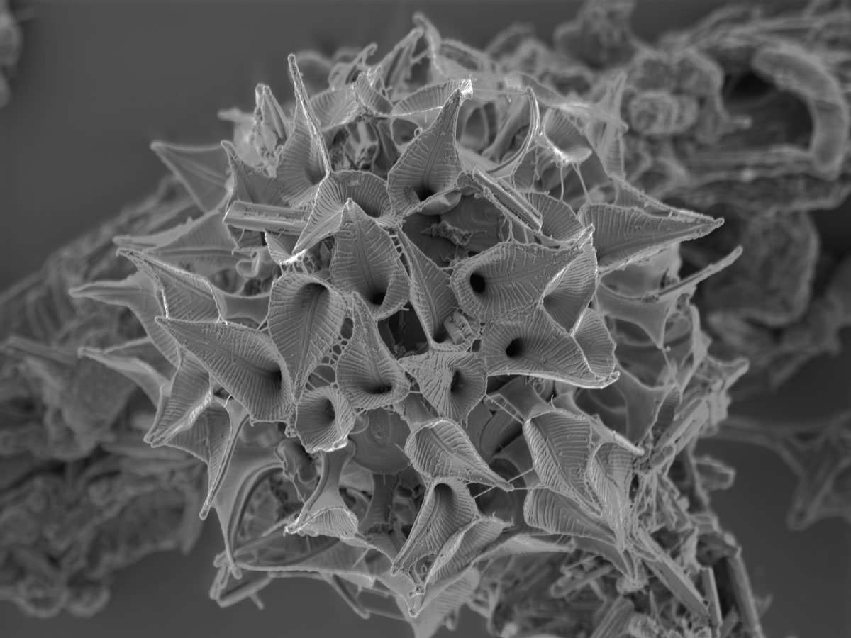
Pterocystis, a very small centrohelid surrounded by spathe-like siliceous scales. (Source: Eckhard Voelcker).

The  website you and Steffen Clauß have created to house these images was named for Eugène Penard, and features a touching biography of the great amoebologist. Do you see your research as a revival or continuation of the kind of work he did? Obviously, alpha taxonomy is less ambitious, these days, and proceeds more slowly; but you have tools at your disposal that can open doors that were closed to pioneers like Leidy and Penard…

I think it would be greatly overstating our capabilities and ambitions to call it a revival or continuation of Penard’s work. But we hope that we can fill a niche. We do groundwork, trying to find amoebae hotspots, and scrutinize them for new species etc.

Next year will be a big change. I have retired from my normal job and Steffen will be working in his normal job only 50%, spending the other 50% in protistology with me. This gives us opportunities to become more professional and to isolate species for further investigation and also for sequencing. When you come home from a normal job, your kids demand attention, the cultures demand attention, there are images that need to be processed, species need to be identified … This has been difficult for us and often when we found something that would require more attention and care we would fail due to time restrictions. Our families have been very supportive and understanding but ultimately, there is only so much you can do when you have a normal job and a family. So now that we made all those changes, we have high hopes for the future.

What is the most challenging hurdle to getting into protists, in your opinion?

Getting into protists is easy, the challenge is coming later: finding a job in protistology that is paying!

Given your professional background in computers and software, do you have any thoughts on how IT could provide more support for protist research?

This is something that constantly amazes me. How can a group of such highly intelligent and educated people live with so little IT infrastructure support? Where is our species database? I want to be able to search based on traits. I want to see images. I want to see the latest papers on this subject. I want to know who has seen it, who is interested in it, etc., etc. I think that protist research would greatly benefit from a certain shared infrastructure. If they all would work for the same institution they would have an infrastructure like this in place within in a short period of time. It would make the entire group so much more efficient and the ROI would be quick and high.

Any other thoughts?

Spend less time surfing the internet and more time in front of your microscope. 😉

I will try to follow that advice!  Thanks for taking the time to do this.

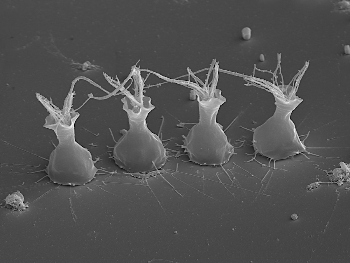
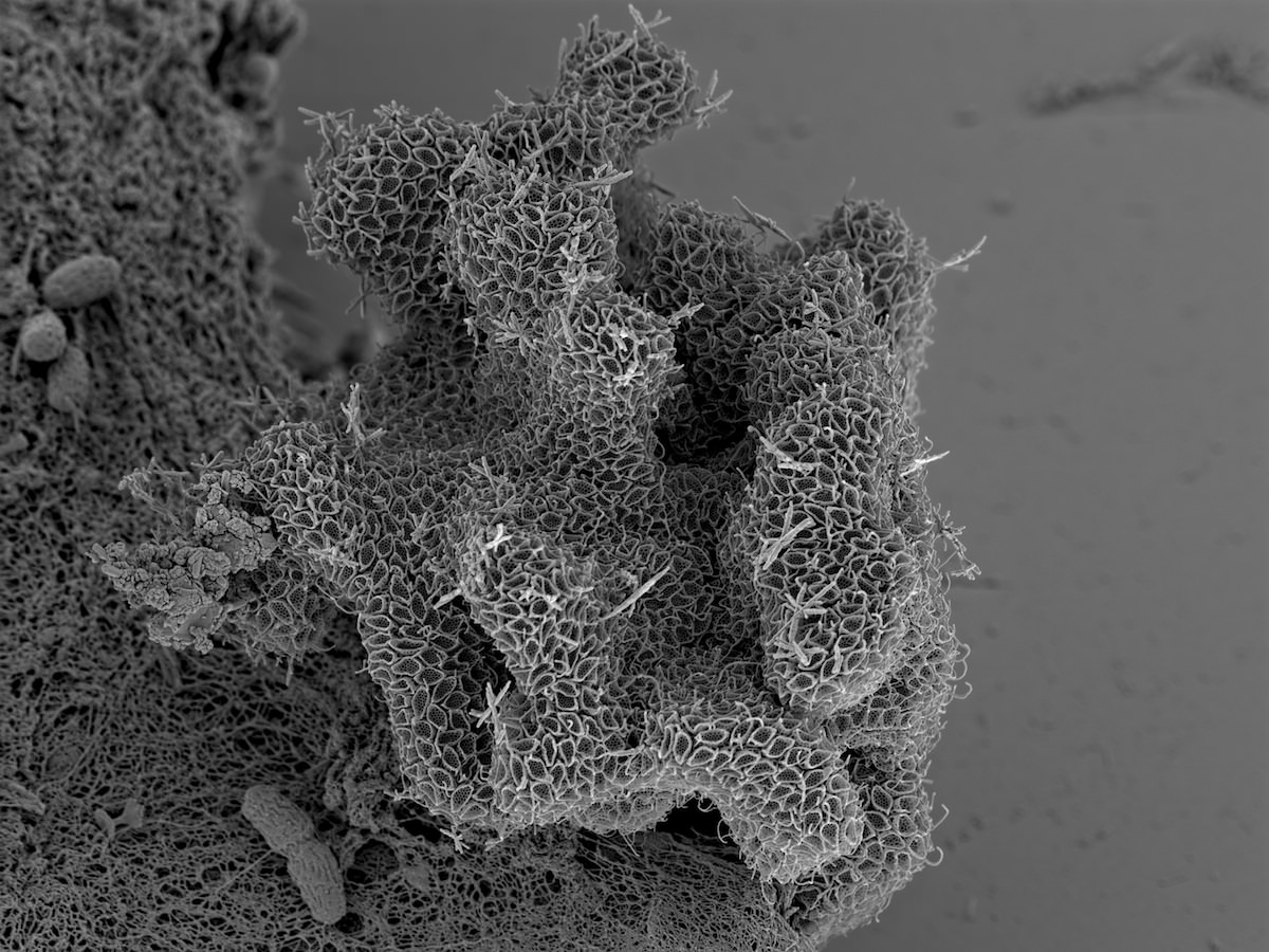
Some choanoflagellates (Choanomonada Kent 1880), members of the protist group most closely related to us. (Source: Eckhard Voelcker)


Dec 112013
 
Ferry Siemensma at his Microscope

Ferry Siemensma at the microscope

Ferry Siemensma is an independent researcher in the Netherlands, and a leading expert on several groups of amoeboid and heliozoan organisms. For the past two years he’s been building a website devoted to his favourite creatures: Microworld: world of Amoeboid Organisms. This was an ambitious project from the beginning, but over time it has gradually turned into something that hasn’t been seen since the comprehensive texts of Cash, Wailes and Hopkinson, or Eugène Penard: a wide-ranging illustrated guide, featuring good formal descriptions, keys for identification, and up-to-the minute taxonomy. Ferry kindly agreed to answer my questions, in the first of what I hope will be a regular series of “Meet the Protistologist” interviews.

Your enthusiasm for your research reminds me of Joseph Leidy’s recorded remark: “How can life be tiresome so long as there is still a new rhizopod undescribed?”   He was not raised in natural science (his father was in the hat business), but even in childhood he was driven “to see how plants and animals are made.” What was your upbringing like?

I grew up in a rural community in the northern part of the Netherlands. My father was a carpenter and I was the oldest of nine children. We always played on our uncle’s farm around the corner, and there I got my first interest in nature. I became a voracious reader of popular books about science and biology and in one of them I read the word “amoeba”. For me it was such a magic word, I don’t know why. From then on I was determined to find such a creature once. I was 14 years old when I bought a cheap Japanese microscope for about $10, which was pretty expensive in 1962. I could recognize some moving protists with it, but no amoebae. Many years later, at the age of 22, I became a teacher in a primary school, where I worked with 11-12 year old children. I also took a study to become a biology teacher and during that study we were offered to buy a Lomo microscope. This Russian instrument was really great, and it had a wonderful 70X water-immersion. From the day I got that scope, my life changed completely!

So, you were a schoolteacher, like Alfred Kahl! What changed in your life after you acquired the Lomo?  

From then on, for more than 12 years, I looked through the microscope nearly every evening. My three daughters were very young at that time and went to bed very early, which was a great advantage ;-). Very soon I got in contact with Rob Wellner, who was looking for a microscope friend. We worked together for two days a week for many years. He taught me a lot, but his main interest was algae and I shifted to amoebae and heliozoa. In that time I got in contact with Dr. De Groot, a well known Dutch amoeba specialist. We smoked a lot of cigars together and had nice talks about our shared interest.

1980-Fred-Page-Cambridge

Frederick C. Page at Cambridge, 1980 (Source: Ferry Siemensma)

When he died in 1981, he left me his scientific literature, with the books of Leidy, Penard and Cash, which was a great luck for me, because in that time it was very difficult to get any papers. There was no internet, which is very hard to imagine now. In the meantime I also got in contact with Fred Page in Cambridge. He is such a pleasant man and we wrote each other a lot of letters. I went twice to Cambridge to see him.

He gave me the address of a Danish microscopist, Niels Willumsen. I phoned him, he invited me and that was the beginning of a very long friendship. One day I got a letter from Rudi Roijackers, who worked at the University of Wageningen. He offered me the use of an electron microscope and that was an excellent chance to get new information about heliozoa. I hunted for heliozoa, isolated them, made dried preparations and once a week we looked at the results in the SEM. We discovered new species and published some papers. I also published two booklets in Dutch about heliozoa and naked amoebae. Than I was invited to write the heliozoan part of the German Protozoenfauna. Fred Page wrote the other part on naked amoebae. The book was published in 1991.

1981-Ferry---Sampling-in-Kortenhoef

Ferry on a sampling excursion, 1981. (Photo by Niels Willumsen, who later capsized in the same boat)

Looking at your publications during this period, you were very active, especially with heliozoans. It must have been a thrill to be among the first to see EM images of the very intricate scales in certain groups! Were there certain organisms that you found particularly fascinating?

1982-Field-study-with-my-Lomo

Ferry in the field with his Lomo, 1982. (Source: Ferry Siemensma)

I was intrigued by the scales of Rabdiophrys species, though they aren’t true heliozoans. Those scales had been studied ten years before by Thomsen and I found them very beautiful subjects to make drawings of. One problem was to interpret the information on the SEM photomicrographs and to form a 3-dimensional image of those scales, which was really a challenge, and very fascinating, which was also true for the base of many kind of spiculae of the true heliozoans.

If you don’t mind my asking, was it a struggle to find funding and free time for this research?  Did you continue to teach?

No, there was no funding, because the work at the university of Wageningen was part of the research of Rudi Roijackers. His interest was Mallamonas and he wanted to find out which scales belongs to heliozoans and which to Mallamonas species. He helped me with the SEM, I helped him with the identification.

Concerning my free time, I did all my research in the evening, the weekends and in my ample holidays. The latter was the big advantage of being a teacher! Schools were closed on Wednesday afternoon, so once a month on Wednesday I jumped into my car and drove to the SEM, one hour driving from my house! Teachers didn’t have a great salary, and they still don’t, so in that time I cleaned every cover glass in order to reuse it 😉

I still clean coverslips, unless they have immersion oil on them!:) In the decade between Nackte Rhizopoda und Heliozoea and the ISOP Illustrated Guide, you seem to have published less frequently. Were you pursuing other interests, or simply working quietly on your own?

1984-Looking-at-Penards-slides-in-the-British-Museum

At the British Museum in 1984, to look at Eugène Penard’s slides. (Source: Ferry SIemensma)

There came a moment that I didn’t find any more new heliozoans. Preparing them for the SEM was a lot of work. First I had to find one, then to decide if it could be a new species, then came the process of removing the cover glass, which was the most complicated part, isolating the specimen, drying it, marking it and finally cutting the slide for the SEM.

After some months without any positive hit, my interest faded. It was at that time, as my girls grew older and needed my attention, that I got very interested in computers and I had a chance to start a science magazine for children – all in my free time. For many years a layer of dust grew slowly on my microscope…

Did you make room for amoebae in your magazine?  It always seems to me that popular science publications have too many telescopes and not enough microscopes!

No, it was intended for 10-14 year old children, and they don’t usually own a microscope.

When did you decide to blow the dust off your microscope, and what brought you back?

Some years ago I got a letter from a woman working at a waterlaboratorium here in the Netherlands. They control the drinkwater quality and had problems with amoebae transporting Legionella bacteria through their filters. They needed my help for identification and had found my name with Google. And there I went to the lab and worked a whole day with a microscope…after so many years. I enjoyed it so much, it was a wake-up call. From that day on, I decided to start again. My first look through my Russian microscope taught me that it was unusable anymore: completely worn out. I bought a secondhand Zeiss standard and later an Orthoplan with DIC and phase contrast and other stuff on eBay. And so I came back home again!

In your website, your experience as an educator and your expertise with amoeboids have come together. How did this project begin?

I started this project about two years ago with the intention to make a survey of all the Dutch amoebae, and as replacement for the book of Hoogenraad & De Groot (1940). Here I could combine my interest in computers, nature, photography and drawing. But soon there came questions from people in the forums who asked for identifications of their findings and I started to publish their photographs also on my site. And then I realized it would be nice to cover the whole world. But it’s a huge project, in fact too much for one person. Every day there are new discoveries to document, new photomicrographs to edit and to archive and new email to answer. But I’m building my site stone by stone… uh.. shell by shell, without hurry and without any pressure. I love it!

While developing your website, you are continuing to do original research.  Can you talk about that work?

1982-Niels-Willumsen-in-his-summerhouse-in-Zweden

Niels Willumsen at his country home in Sweden, in 1982. (Source: Ferry Siemensma)

I’m very interested in the variation within populations. Probably you know how difficult it is to distinguish between certain Centropyxis species, and the same is true for Difflugia, Euglypha, Cyphoderia and many other taxa. I’m measuring populations from different localities and will see what comes out. At the moment I have an Austrian sample with a population of Pseudonebela africana, which is new for Europe. It has been described from Africa and Brazil as having dimensions between 78 and 100 µm, but I find shells which are 85-185 µm long. That’s really interesting, and shows how little we know of populations in the wild. As these specimens are living, I also have the opportunity to observe their feeding behavior which is the same of that of Difflugia rubescens, and these specimens also have the same red color. They must be related. I’m fascinated by the behavior of amoeboids. I’m using a nice and simple, but very successful method for observations, the so-called “wet chambers” where I keep my wet mounts. It’s always a surprise, what comes up in such mounts after several days. Not seldom some species multiply rapidly and attach to the cover glass. which is excellent for observations. Mmm… I can go on for many hours…

It would be a pleasure to spend those hours with you, and I hope I will have the chance, one day.  Thank you so much for taking the time to do this.

Dec 082013
 

I’ve added a number to the title of this post, because I expect to make “Protist Homes” a regular feature. I had intended it to be an idea-free zone, devoted to uncomplicated wonderment (kind of like the tours of celebrity homes and stately residences on HGTV).  But I know some ideas and research will creep in, because it’s hard to feel wonderment without actually starting to wonder.

Difflugia acuminata

Difflugia acuminata, from a pond in Wakefield, Quebec


Consider the little shell in the image to the left.  Barely two tenths of a millimetre long, it was constructed by an amoeboid, using found materials: tiny grains of sand, glued together with some sort of proteinaceous cement.

How does a single-celled amoeba build such a perfect bottle, from such randomly-shaped material?  And why did my grade-school macaroni art projects turn out so much worse?


The organism that once lived in that test is dead (testate amoebae cannot leave their shells without dying). In life,  it would have looked and behaved like the one in this video, by my fellow protist-watcher, Francisco Pujante:

All members of the genus Difflugia build their shells–or tests, as they’re properly calledout of stuff they find lying around, such as grains of quartz or discarded diatom shells, binding them into a matrix of organic secretions. It turns out that they are quite selective about the particles they use for this work, and researchers have gone to a lot of trouble to figure out what criteria amoebae use when picking their building materials.

Difflugia 3D rendering 2

3D rendering of Difflugia oblonga, created by Châtelet et al. Click image to show source.

Recently, a group in France went so far as to create complete three-dimensional reconstructions of two specimens of Difflugia oblonga, using a process called x-ray microtomography. They calculated the volume, shape and orientation of each grain in their specimen tests, and gathered information about the density and composition of the individual particles. Yes, you read that correctly: they analyzed and modelled every micro-sandgrain in amoeba shells that are themselves smaller than the grains of sand you might find on an ordinary beach.

Using all this data, they compared the composition of the tests to the natural sediments in the ponds where they were collected. From their analysis, it appears that these amoebae really are selecting the smaller grains, and also showing a taste for certain minerals (quartz is preferred, while the more abundant calcite is shunned). What’s more, the amoebae seem to be selecting materials with a consistent density, which the authors speculate is to ensure the test will be perfectly balanced!

Difflugia grain sizes


The paper (du Châtelet et al, 2013), includes a handy graph, comparing the sizes of each of grain of sand in two Difflugia tests to a distribution plot of the size of sand particles in the surrounding pond sediment.


Building with quartz is fine, if you are lucky enough to live in the mud bottom of a pond where chunks of silicate minerals are available.  But what if your habitat is a floating clump of algae in a mineral-poor spagnum pool?  There, the best source of silicate particles might be the siliceous shells (frustules) of your fellow protists the diatoms, which have the ability to build their little glass canoes from dissolved silica (silicic acid) found in the water around them. The testate amoeba Difflugia baccillariarum–typically found in sphagnum pools, where sand is in short supply–has figured out how to glue diatoms around itself as a means of protection.  I find these guys, sometimes, in samples from the Mer Bleue bog,  and always stop to look at them.  They are truly amazing:

It is hard not to admire this crafty amoeboid, building its home from the bodies of other organisms.  Sitting here in my post-and-beam house, assembled from the trunks and branches of felled trees, I feel a certain kinship.