Jul 112016
 

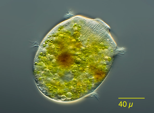
There’s a natural pond down the road, where I collect samples. I always go to the same spot, the edge of a shallow basin in the “littoral zone”, where the pond shades into the shore. I follow the same routine each time: I dip a turkey baster into the soft muck–organic sediments enriched with duckshit and decomposing leaves–and suck out a few cc’s for the bottom of the jar; then I ladle a bit of surface water over the mud, to create a miniature approximation of the natural site.

Turtle Pond2

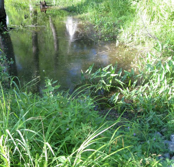
Sampling Site in Turtle Pond

I’ve taken hundreds of samples there over the years, from an area no bigger than a coffee table.  And, despite having done this regularly for about half a decade, nearly every jar I bring home contains something I’ve never seen before.

Of course, to a microbe, a coffee table-sized site is actually quite big. If you were a typical one-celled protist–say, 50 micrometres from tip to tail–a four-foot patch of pond would be more than 24,000 times longer than your body. From a microbe’s point of view, then, my sampling basin is equivalent to a lake 28 miles long. Scaled up to human size, the entire pond would be at least 1,350 miles across, with a greater surface area than the Mediterranean Sea! So, there’s quite a lot of available habitat in the small body of water I call “Turtle Pond.”

At any given time, only a small fraction of the site’s potential diversity will be visible. The mix of species can change from one hour to the next, as weather and random events alter water chemistry, light levels, temperature and organic nutrients. A few hours of sunlight might kick off a sudden bloom of cyanobacteria, soon followed by nassulid ciliates that eat strands of blue algae like spaghetti; then, a few minutes of rain disperse the bloom, the nassulids disappear, and are replaced by some other combination of species. The new populations have barely established themselves, when a turtle (which, from a microbe’s point of view, is about 7 miles long), blunders into the shallows, churning up mud, mixing oxygen-rich upper layers with the anoxic sediments below, and everything changes again. Each shift in the pond creates new opportunities for some organisms, and makes life impossible for others. One species blooms, and another goes into dormancy, withdrawing into resting cysts or simply lurking in reduced numbers, waiting for better days.

The microbial “seed bank” can store a staggering diversity of such hidden opportunists, which emerge from their cysts only when conditions are exactly right. Some are “weed” species, generalists that crop up in nearly every sample. Others have very particular needs, and rarely appear. A few may never find what they need to grow and reproduce. If any cysts of salt-loving (halophilic) protists should blow in from some distant shore, they are probably out of luck.

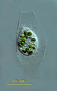
Recently, I brought home a few jars from the usual spot, and found three rather flamboyant species to cross of my life list. One was Actinobolina radians, an oddball I’ve been hoping to find for years (ever since I first saw it’s mugshot in Vladimir Schewiakoff’s beautifully-illustrated Infusoria Aspirotricha of 1889). Actinobolina is a ciliate in the class Litostomatea, a group of rapacious and morphologically hilarious carnivores. The class includes such celebrity ciliates as the improbably elastic Lacrymaria, the elephant-nosed Dileptus, and a frisky little keg full of death called Didinium, which eats other ciliates like popcorn. Like its cousins, Actinobolina is a hunter. However, it has a unique strategy for capturing prey. When swimming about, it looks much like any other ciliate: egg-shaped, and uniformly covered with cilia. Once it has a found a suitable feeding site, it comes to a stop and slowly extends a large number of long, stiff tentacles, each of which carries toxic extrusomes (cellular darts) to be released on contact into its victim’s body. In this state, it looks rather like a heliozoan “sun animalcule”.   With its tentacles extended, Actinobolina lies in wait like a spider in its web, until some tasty creature–a rotifer , or another ciliate–bumps into it. The toxic extrusomes are deployed and the victim is paralyzed, whereupon the ciliate withdraws its tentacles, pulling the stunned prey toward its cellular mouth.

Unfortunately, I didn’t manage to record a successful hunt, but I did get some footage of the ciliate extending and retracting its tentacles.  And really, I was just pleased to have seen this bizarre organism.

Another creature that appeared in my jars was the odd, oxygen-hating amoebozoan Pelomyxa, an indiscriminately hungry thing that looks like a tumbling bag full of rocks and food. I see Pelomyxa fairly often, but this one was a different species, smaller and more colorful, and with a distinctive style of movement, featuring frequent lateral “eruptions” of pseudopods. I believe it is Pelomyxa binucleata, a species first identified in the 19th century. For a time, in the late 20th century, it was believed that P. binucleata and all “species” of Pelomyxa, were really just forms and phases of a single species, Pelomyxa palustris. However, since 2004 the Russian researcher Alexander O. Frolov and his collaborators have been repopulating the genus, discovering new species and rehabilitating older ones. There are more than half a dozen, now.

Pelomyxa is also interesting because of its role in the rise and fall of the fabled Lost Protist Kingdom, once known as Archezoa. At one time, in the 1980s and early 90s, Pelomyxa was believed to be a primitive eukaryote, a “living relic of the Proterozoic era” (Margulis & Sagan, 1990) lacking such basic organelles as mitochondria, centrioles, flagella/cilia, endoplasmic reticulum, Golgi bodies and even chromosomes (Margulis & Chapman, Kingdoms and Domains, 2009). It was seen as sort of a missing link between eukaryotes and their bacterial predecessors, and was placed along with an assortment of other anaerobes, in the short-lived Kingdom Archezoa. However, that hypothesis began unraveling within just a few years. Pelomyxa were found to have centrioles and flagella after all, as well as mitochondrial remnants (“mitochondria-derived organelles”). In fact, they turned out to possess most of the stuff you’d expect to find in a eukaryotic cell, along with some nifty new adaptations to anaerobic living, so there was really no reason to regard them as “primitive” at all. When genetic analysis of the genus was finally done, it was placed squarely within the phylum Amoebozoa.  Archezoa itself–briefly a great Kingdom, ruled by a benign despot named Thomas Cavalier-Smith–was quietly removed to the museum of obsolete evolutionary hypotheses.

Finally, my samples turned up a single specimen of Supraspathidium, a rather long haptorid ciliate with wide, blubbery lips. I’m afraid this guy will be of no interest to anyone but the most devout ciliatophile, but I’m posting it anyway because it’s my blog. Supraspathidium resembles ciliates of the better-known genus Spathidium, except that it has numerous contractile vacuoles, instead of a single big one in the posterior of the cell. This one has a long, wormy macronucleus, and generally resembles a spathidiid described by Eugène Penard in 1922,under the name Spathidium vermiforme.

REFERENCES

Feb 182015
 

In my previous post on the lorica-dwelling scuticociliate Calyptotricha pleuronemoides, I mentioned that only two substantial articles have been written about the species since its discovery (apart from brief descriptions in various places). Until yesterday, I’d been unable to find the second article, which appeared in the German microscopy journal Mikrokosmos in 1999. Luckily, one of the co-authors, Martin Kreutz, was kind of enough to send me a copy!

calyptotrichapleuronemoides1_mkw

Calyptotricha pleuronemoides. Image by Martin Kreutz. Source: micro*scope. Click on image for link to source.

Martin tells me he sees the organism frequently in water from the sphagnum ponds of Simmelried, a system of bog lakes, like the one in Ottawa’s Mer Bleue where I sometimes find Calyptotricha. He and Philipp Mayer provide a good redescription of the ciliate, with morphometrics. Their measurements match those of the specimens I’ve found, and agree with those of Phillips and D.S. Kellicott. (All sources give a size range somewhat smaller than that recorded by Alfred Kahl, who gives 50 µm for the length of the cell, and 85 µm for the lorica).

The Mikrokosmos article is difficult to find, so I thought I might give a brief redescription of the species, based on the information collected by Kreutz and Mayer:

Calyptotricha pleuronematoides:  Pleuronematid ciliates, in spindle-shaped hyaline lorica 56-75 µm long, tubular with narrowed openings at either end, 9-13 µm in width. Lorica broadens in the middle to a width of 23-24 µm. Cell body resembling Cyclidium, slightly flattened back to front, 22-35 µm long, 15-24 µm wide, somewhat concave on the ventral surface, where oral apparatus occupies 3/4 of body length. L-shaped undulating membrane, made up of fused cilia, 14 µm long. Caudal cilium 10-12 µm long; roughly 17 somatic kineties, spaced 2-3 µm apart. Most specimens with 8-15 zoochlorellae, colorless examples rare. Single oval macronucleus, with small spherical micronucleus. CV in posterior.

The authors mention that specimens are seldom seen outside of their loricas. Free-swimming individuals move quickly, but not as jerkily as Cyclidium.  I happen to have recorded a free-swimming Calyptotricha, last year, so I might as well post it here:

References:

Kreutz, Martin, and Philipp Mayer. “Artikel-Calyptotricha pleuronemoides-Ein Ciliat in einer Rohre.” Mikrokosmos 88.1 (1999): 27-30.
Feb 132015
 

A few years ago, I looked in a sample of water from a bog lake, and saw something like a hyperactive avocado shifting around inside in a tiny kerosene lamp:

The architect of that pretty dwelling is the ciliate Calyptotricha pleuronemoides. The species and genus were discovered in 1882, in samples from a pond near Hertford, England, by an amateur naturalist named Frederick W. Phillips.  Not much is known about him.  During the 1880s, he was an active member of the Hertfordshire Natural History Society and Field Club, to whom he occasionally read essays on “The Protozoa of Hertfordshire,” based largely on the classification scheme in William Saville Kent’s Manual of the Infusoria.  He was a Fellow of the Linnean Society of London, and he found and named a few new taxa.

In his very first glimpse of the creature, Phillips was lucky enough to catch it in the act of building its lorica. “At first sight,” he writes, “I thought it was an embryonic or encysted stage of some monad; but upon applying a magnifying power of some 900 diameters, I observed that it possessed a singular vibratile membrane, closely resembling that which characterizes the members of the family Pleuronemidae.” A week later, Phillips looked at it again, and discovered that “the lorica had increased in size, and that one end was elongated into a teat-like form.” At this stage, he accidentally allowed the sample to dry out, leaving the organism’s empty, half-finished lorica still attached to a strand of pond-weed. He made a nice drawing of what he’d seen.

Calyptotricha pleuronemoides from Phillips resized

A. First stage B. The same, further developed C. End view of lorica D. The perfect animal E. Ventral view (adapted from Phillips)

To modern readers, accustomed to the impersonal, passive style of scientific writing–“samples were collected,” “living cells were isolated and observed”–there is something pleasingly candid about the way Victorian naturalists report their findings. Phillips doesn’t just describe his new genus, he spins us the tale of its discovery, including the mishap that destroyed his first specimen, and his initial misreading of the oval shell, after which he takes us to the very moment of discovery when he exposed the creature’s true nature by “applying a magnification of 900 diameters.” Something about that reminds me of the exploration literature of the same period. It’s probably not just an accident of style: Victorian microscopists were explorers. Superior lenses and stains had opened up a miniature Dark Continent on their laboratory benches, and a gentleman adventurer from somewhere like Hertfordshire could now penetrate these hidden realms, returning with breathless accounts of what he had seen. A session at the microscope was an expedition into the unknown.

In our time, researchers are expected to pile up some data before going to print, and nobody would attempt to erect a new ciliate genus on the basis of a brief observation of a few specimens. No doubt that is a good thing: the 19th century left a big legacy of poorly defined taxa, many of which are still desperately in need of revision.  But this kind of field work, as sketchy and dilettantish as it might seem now, has largely been put to one side without really being replaced by anything better.  Outside of a few centers of activity, ciliate field work has slowed to a crawl.  Consider the fact that 132 years after Phillips wrote his three-page note on Calyptotricha pleuronemoides it is still one of only two substantial treatments of the species, and the only source that describes the construction of its curious lorica.  Anyone who wants to know more about this ciliate than its name, has to travel back to the 19th century.

poke bonnet

A straw poke-bonnet, from the early 19th century. (Click for source)

Needless to say, the old information is not always reliable.

Phillips perceived immediately, and rightly, that Calyptotricha is closely related to the more common ciliate Pleuronema. Like its cousin, it is equipped with a large, billowing membrane that runs along the right side of its oral aperture. However, Phillips badly misunderstood the shape of this structure, describing it as “a membranous trap, or velum, which in form resembled the old-fashioned poke-bonnet.”

When I first read that passage, the comparison to a “poke-bonnet” confused me. The undulating membrane of pleuronematid ciliates is shaped something like a sail, or a flag: a sheet of fused cilia running along one side of the organism’s mouth. Phillips, however, interpreted this structure (which, admittedly, is very difficult to see clearly in the light microscope) as a sort of hood or canopy covering the oral aperture of the ciliate. If you look closely at his illustration, you can see that he has drawn it as a baggy tube.

Calyptotricha's undulating membrane resembles a sail or banner (image adapted from Colin R. Curds, British and Other Freshwater Ciliated Protozoa)

The true shape of Calyptotricha’s undulating membrane (image from Colin R. Curds, British and Other Freshwater Ciliated Protozoa, with arrows added)

Evidently, it was this imagined resemblance to a poke-bonnet that prompted him to give the genus its curious name, Calyptotricha, constructed from the Greek calyptos (“veiled” or “covered”) and trich (“hair”). It seems the “haired” holotrichous ciliate reminded him of a woman’s head, on top which the membrane sits like an old-fashioned hat!

It’s an example of how expectation shapes observation. In interpreting this membrane as an enclosed hood, he was deferring to an earlier error by his illustrious contemporary William Saville Kent. Writing about Pleuronema, Kent says: “[T]his membranous trap may be appropriately compared with the extensile hood of a carriage or an outside windowshade forming, when expanded, a capacious hood-shaped awning, and when not in use being packed away in neat folds close around the animalcule’s mouth.”

The “extensile hood” Kent mentions was a common convenience on carriages of his day, and provided a compelling mechanical analogy for the “neat folds” with which he imagined Pleuronema pulled back its velum.

Barouche image 2Here, for comparison, is Kent’s illustration of Pleuronema chrysalis, which I’ve inverted to showcase its “extensile hood.”

Pleuronema chrysalis, from W. S. Kent's A Manual of Infusoria. Put wheels on it, and you have a chuck wagon.

With wheels, it would make a good chuck wagon.

To modern workers familiar with the morphology of hymenostome ciliates, as revealed in specimens that have been stained with silver, this is an implausible design. However, to Kent, who had done pioneering work on choanoflagellates, it seemed reasonable to speculate that Pleuronema’s hood might share “a distant homological relationship” with the “delicate funnel-shaped membranes” found in the collared flagellates, which really do wear something a bit like a straw poke-bonnet (but on the back end of the cell).

Finally, since we’ve been talking about Pleuronema and her sisters, I’ll post some footage of one, quietly browsing on bacteria in water taken from a tidal pool on the coast of Maine:

REFERENCES

Nov 182013
 

I used to study English Literature.  I did that for a decade and a half, at two universities, and eventually wrote a doctoral thesis on “Postwar American Poetry.”  In all the years I spent researching poetry,  nobody ever asked me why I would want to study something like that.  It’s not that everybody loves poetry. Most people, in my experience, really cherish the time they don’t spend reading poetry. But some people do like it well enough, and we all know that people of that kind are out there, somewhere.

Protists are different.  Apparently, if you happen to be interested in those, you have some explaining to do.  In fact, it’s the first thing most people ask, when I tell them how I’ve been spending my days: Why are you interested in that?

It always deflates me a bit, which is fine, I would not want to be too puffy.  But would they ask me that if I were studying…tigers?

wcs_tigerresearcher

John Goodrich, Tiger Researcher (Image: A. Rybin.  Click to see source)

I’m pretty sure that guy never has to explain why he loves what he does. He catches live tigers in the wild and cuddles their babies!   That’s a solid 40 megafonzies on the Cool-O-meter. But when I mention that I’m interested — very, very interested — in “protists,” I can tell that some people do not necessarily think it is a good thing.

Part of the problem is the word “protist” itself. From a public-relations point of view, it is a mess. First, it sounds too much like a certain other English word. When the subject of protists comes up (as it always does, if I can work it into the conversation) people often mishear me, and think that I like to study and observe “protests.” Which is not so implausible…I’m sure there are “protest watchers” out there, as there are “storm chasers” and “chicken hypnotizers.”   But even when I spell the word out, people over a certain age (my generation, that is)  don’t recognize it anyway. We grew up calling these bugs either “algae” (the placid green ones that remind us of plants) or “protozoa” (the colourless ones that boogie around and try to eat each other, like miniature animals).

These days, the word “protozoa” is being phased out, in professional circles. The reason for this is perfectly sound. It means “first animals,” and protists are not animals of any kind.  The mushrooms in your fridge are more closely related to you (and your dog, and your dog’s fleas), than any of the protists in your koi pond. But although both words were coined in the 19th century, the word “protozoa” is the one that caught on and stuck. In marketing terms, it has good “brand awareness.” So, here we have a field, protozoology, that was already on the margins of the public mind, which now goes by a “new” and unfamiliar name.

And finally, once we’ve established what it is we are talking about, some people still fail to see–amazing as it sounds!–why a person might care about such a thing.  Even biologists, who should know better, tend to think of protists as a weird little sideshow in the circus of life, where the big tent is reserved for elephants, horses and (needless to say) tigers.

I’ll talk about that in my next post, I think.

First, though, since I’ve been belabouring the word “protist,” what exactly do we mean by it?  This blog is about protistology, so there will be ample time to explore that in depth, but a quick working definition might be useful. If I may adapt the excellent and succinct definition offered by Psi Wavefunction, a protist is any organism that is not a bacterium, not a fungus, not a plant, and not an animal. Which means that protistology is, effectively, the study of organisms that nobody else cares about.

Any eukaryote that is not a plant, an animal, or a fungus. – See more at: http://skepticwonder.fieldofscience.com/2008/11/what-is-protist-eukaryotic-tree-of-life.html#sthash.Yup6wXZj.dpuf
Nov 142013
 

When I was eight years old, I knew what I wanted to be when I grew up.

I wanted to be this guy:

Forty-five years later, I finally look a bit like that. It’s not the wild hair and bulging eyes (which I’ve always had): it’s the glassware. At 53, I finally have an erlenmeyer flask!  I also have a box of pipettes, and a big fat falcon tube filled with a yellow fluid that might well turn into a powerful explosive if I ever let it dry out.  I do not have a brain in a jar, but it’s only a matter of time.

In short: at a fairly late stage of life I am trying to become, in my own small way, a kind of scientist.    I have no scientific training at all.   I am a poet, who happens to have written a bit about protists and microscopy.    I’m hoping this blog will give me a place to write about the slow and sometimes awkward process of learning to think scientifically.

What I’m beginning here could be described as a “citizen science”  blog, but that term doesn’t sit well with my inner eight-year-old.   “Citizen science” is all about “making a contribution,” like those diligent bird-counters, reporting their sightings to the Cornell Lab of Ornithology; or telescope fanciers up till all hours, sifting the barely-perceptible specks from the almost-invisible flecks.   There’s nothing wrong with doing your bit for the cause, but that is not what pulled me into this.  (And really, can you picture a power-hungry “citizen scientist” cackling over a fuming flask?)

Since we do have to distinguish the self-taught amateur from the qualified pro (if only to decide who gets a turn at the electron microscope), I prefer the term “outsider science,” which hints at the kind of not-altogether-healthy obsessiveness that drives a person like me.   Of course, that epithet is, if anything, even less flattering.  While the “citizen scientist” may be, at worst, a gormless do-gooder, the “outsider scientist” appears to be (in the journalistic imagination, at least) a flat-out crank:


(Image: Reidar Hahn, via Symmetry Magazine)

But if the “outsider scientist” is a kook, he is at least a passionate one; and I’m not too proud to admit that something in that picture kind of reminds me of me.

Also, protistology is itself something of an “outsider science,” which has a long history of providing an intellectual home for dedicated autodidacts.  I will talk a bit about that in my next post, or the one after that.